The mammalian brain is a complex and essential organ involving complex processes like memory, cognition, and feeling, and is linked to various mental diseases. However, our knowledge and research of this part are limited, and the research on the brain will be a long-term exploration. Currently, the brain imaging of living animals can intuitively display the morphological changes in the brain during life activities, allowing researchers to study the functional activities of the brain, which is vital for understanding the structure and function of the brain. Owing to the thick brain tissue, brain imaging in vivo is affected by factors such as light scattering, light absorption, and aberration, which hinders the optical imaging of the deep structure of the brain. Similarly, the resolution of the general optical microscope cannot exceed 200 nm due to an optical diffraction limit, and it cannot distinguish the subtle structures such as neuronal synapses in the brain nor analyze the structures that play a vital role in brain functional activities.
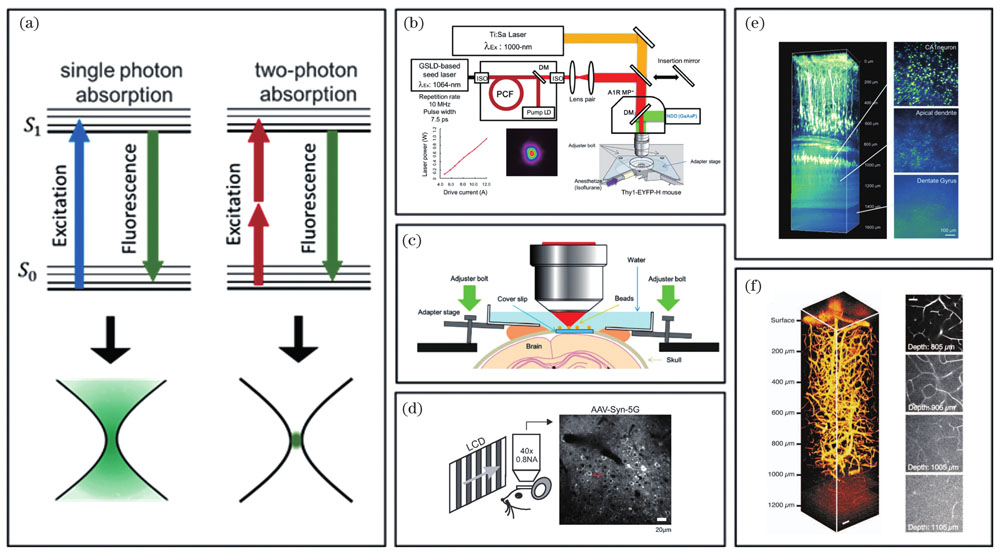
The multiphoton imaging technology was used in the past to improve imaging depth. This technology uses a nonlinear excitation effect and often utilizes near-infrared excitation, which can stimulate short-wavelength fluorescence signals with long-wavelength lasers. With low scattering and the absorption of the near-infrared light of the tissue, it can attain a high penetration depth and reduce the background interference of the tissue. It is an important method for thick tissue imaging. Additionally, multiphoton imaging only stimulates fluorescence at the focal plane, which has the natural characteristic of "light slicing" , which decreases the signal interference of the defocus plane and tissue stimulation damage. Therefore, the multiphoton imaging technology is suitable for imaging the brain in vivo. Nevertheless, the diffraction limit remains a major concern for this technology.
Introducing the super-resolution imaging technology makes nano-optical imaging possible. The super-resolution microscopic imaging technology is applied to the in vivo imaging of the brain, which can dictate the delicate features like neuronal synapses and then study the functional activities of the brain at the in vivo level. Meanwhile, based on the traditional multiphoton imaging technology, introducing the super-resolution microscopic imaging technology, combined with the benefits of penetration depth and spatial resolution, can enhance the in vivo imaging ability of the brain, which is beneficial in the fields such as neuroscience and brain disease research.
First, the super-resolution imaging technologies used in brain imaging, stimulated emission depletion (STED) and structure illumination microscopy (SIM), are summarized (Table 1). Currently, the STED imaging technology is the main living animal brain imaging technology (Fig. 2). Hell’s research group applied STED to the imaging of neuronal dendritic spines or actin filaments in the mouse cerebral cortex and achieved a lateral resolution exceeding 70 nm. However, there are few reported SIM imaging technologies. Although the spatial resolution of SIM is essentially limited to 100 nm, its excellent imaging depth also has potential. Additionally, both super-resolution technologies have been tried to combine with the two-photon imaging technology. Although they have improved the imaging depth to a certain extent, the depth has not exceeded 120 nm, which does not achieve the desired effect of two-photon imaging. Moreover, according to the research status, the main challenges faced by the super-resolution imaging technology in living brain imaging are analyzed and discussed, and the five aspects of imaging speed, imaging depth, imaging time, fluorescent probe, and animal state are summarized. Furthermore, the application of the super-resolution imaging technology still needs to make efforts in spatial resolution. Finally, suggestions for solving these problems are presented.
The super-resolution imaging technology can break through the optical diffraction limit and be applied to living brain imaging. It is of great importance to study the structure and function of the brain at the nano level. Presently, STED and SIM are two main application technologies, and good progress has been made in combination with two-photon imaging. Presently, applying the super-resolution microscopic imaging technology in living brain imaging is not common, and it still faces many challenges. To make the best use of the super-resolution imaging technology, appropriate imaging technology, fluorescent probe, and experimental object should be selected based on the experiment requirements, and the imaging depth, imaging speed, and imaging time should be stable. The super-resolution microscopic imaging technology has great potential and its application and development will enhance the research of brain imaging in living animals.