• Acta Optica Sinica
  • Vol. 39, Issue 2, 0211002 (2019)
Tingyue Zheng1、*, Chen Tang1、*, and Zhenkun Lei2
Author Affiliations
  • 1 School of Electrical and Information Engineering, Tianjin University, Tianjin 300072, China
  • 2 State Key Laboratory of Structural Analysis for Industrial Equipment, Dalian University of Technology, Dalian, Liaoning 116024, China
  • show less
    DOI: 10.3788/AOS201939.0211002 Cite this Article Set citation alerts
    Tingyue Zheng, Chen Tang, Zhenkun Lei. Multi-Scale Retinal Vessel Segmentation Based on Fully Convolutional Neural Network[J]. Acta Optica Sinica, 2019, 39(2): 0211002 Copy Citation Text show less
    Structural diagram of residual block
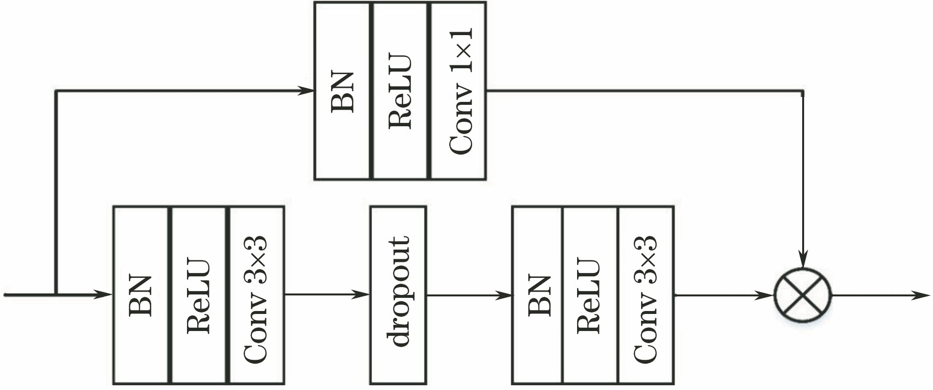
    Fig. 1. Structural diagram of residual block
    Schematic of multi-scale ASPP module
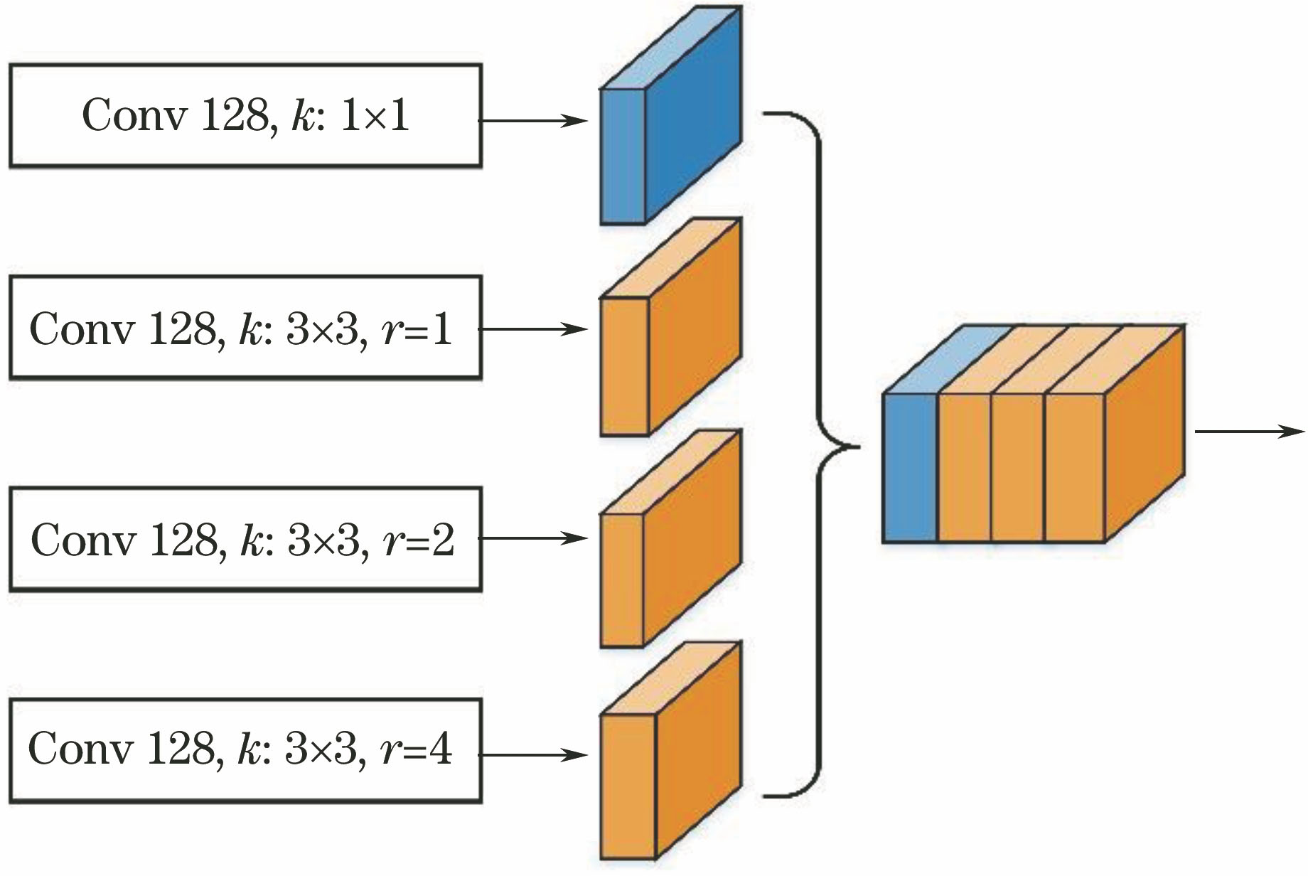
    Fig. 2. Schematic of multi-scale ASPP module
    Structural diagram of network for retinal vessel segmentation
    Fig. 3. Structural diagram of network for retinal vessel segmentation
    Image preprocessing. (a) Typical fundus image of DRIVE dataset; (b) fundus image after preprocessing
    Fig. 4. Image preprocessing. (a) Typical fundus image of DRIVE dataset; (b) fundus image after preprocessing
    Segmentation test results based on DRIVE dataset. (a) Original fundus images; (b) segmentation standard images; (c) segmentation results of images
    Fig. 5. Segmentation test results based on DRIVE dataset. (a) Original fundus images; (b) segmentation standard images; (c) segmentation results of images
    Segmentation test results based on STARE dataset. (a) Original fundus images; (b) segmentation standard images; (c) segmentation results of images
    Fig. 6. Segmentation test results based on STARE dataset. (a) Original fundus images; (b) segmentation standard images; (c) segmentation results of images
    Segmentation results in local areas. (a)(b) Original fundus images; (c)-(f) local fundus images; (g)-(j) segmentation standard images; (k)-(n) segmentation results of images
    Fig. 7. Segmentation results in local areas. (a)(b) Original fundus images; (c)-(f) local fundus images; (g)-(j) segmentation standard images; (k)-(n) segmentation results of images
    DatasetMethodRSe /%RSp /%RAcc /%RAUC /%
    DRIVE2nd human observer77.6097.2494.72
    Proposed method80.5397.6795.4697.71
    STARE2nd human observer89.5293.8493.49
    Proposed method82.9997.9496.8498.17
    Table 1. Average performance evaluation results based on DRIVE and STARE datasets
    TypeMethodYearRSe /%RSp /%RAcc /%RAUC /%
    Ref.[3]201174.1097.5194.34
    UnsupervisedRef. [5]201462.8098.4093.80
    methodsRef. [2]201576.5597.0494.4296.14
    Ref. [7]201574.2098.2095.4086.20
    Ref. [9]201274.0698.0794.8097.47
    Ref. [13]201581.7397.3397.6794.75
    SupervisedRef. [12]201677.6397.6894.9597.20
    methodsRef. [14]201675.6998.1695.2797.38
    Ref. [15]201676.0395.23
    Ref. [17]201775.0197.9594.99
    Proposed method201880.5397.6795.4697.71
    Table 2. Performance comparison of the proposed and other methods based on DRIVE dataset
    TypeMethodsYearRSe /%RSp /%RAcc /%RAUC /%
    Ref. [3]201172.6097.5694.97
    UnsupervisedRef. [5]201458.6098.7094.48
    methodsRef. [2]201577.1697.0195.6394.97
    Ref. [7]201578.0097.8095.6087.40
    Ref. [9]201275.4897.6395.3497.68
    Ref. [13]201581.0497.9198.1397.51
    SupervisedRef. [12]201678.6797.5495.6697.85
    methodsRef. [14]201677.2698.4496.2898.79
    Ref. [15]201674.1295.85
    Proposed method201882.9997.9496.8498.17
    Table 3. Performance comparison of the proposed and other methods based on STARE dataset
    Tingyue Zheng, Chen Tang, Zhenkun Lei. Multi-Scale Retinal Vessel Segmentation Based on Fully Convolutional Neural Network[J]. Acta Optica Sinica, 2019, 39(2): 0211002
    Download Citation