• High Power Laser and Particle Beams
  • Vol. 34, Issue 12, 122001 (2022)
Tiankui Zhang1, Lianqiang Shan1, Minghai Yu1, Feng Lu1, Weimin Zhou1、*, Chao Tian1, Fang Tan1, Yonghong Yan1, Feng Zhang1, Zongqiang Yuan1, Qiuyue Xu1, Weiwu Wang1, Zhigang Deng1, Jian Teng1, Dongxiao Liu1, Lei Yang1, Wei Fan1, Yue Yang1, Kainan Zhou1, Jingqin Su1, Yuchi Wu1, Yongkun Ding2, and Yuqiu Gu1、*
Author Affiliations
  • 1Laser Fusion Research Center, CAEP, P. O. Box 919-986-6, Mianyang 621900, China
  • 2Institute of Applied Physics and Computational Mathematics, Beijing 100088, China
  • show less
    DOI: 10.11884/HPLPB202234.220186 Cite this Article
    Tiankui Zhang, Lianqiang Shan, Minghai Yu, Feng Lu, Weimin Zhou, Chao Tian, Fang Tan, Yonghong Yan, Feng Zhang, Zongqiang Yuan, Qiuyue Xu, Weiwu Wang, Zhigang Deng, Jian Teng, Dongxiao Liu, Lei Yang, Wei Fan, Yue Yang, Kainan Zhou, Jingqin Su, Yuchi Wu, Yongkun Ding, Yuqiu Gu. Source-coded radiography technique with high spatial-resolution for X-ray source driven by ps-laser[J]. High Power Laser and Particle Beams, 2022, 34(12): 122001 Copy Citation Text show less
    Diagram of laser driven X-ray source coding radiography
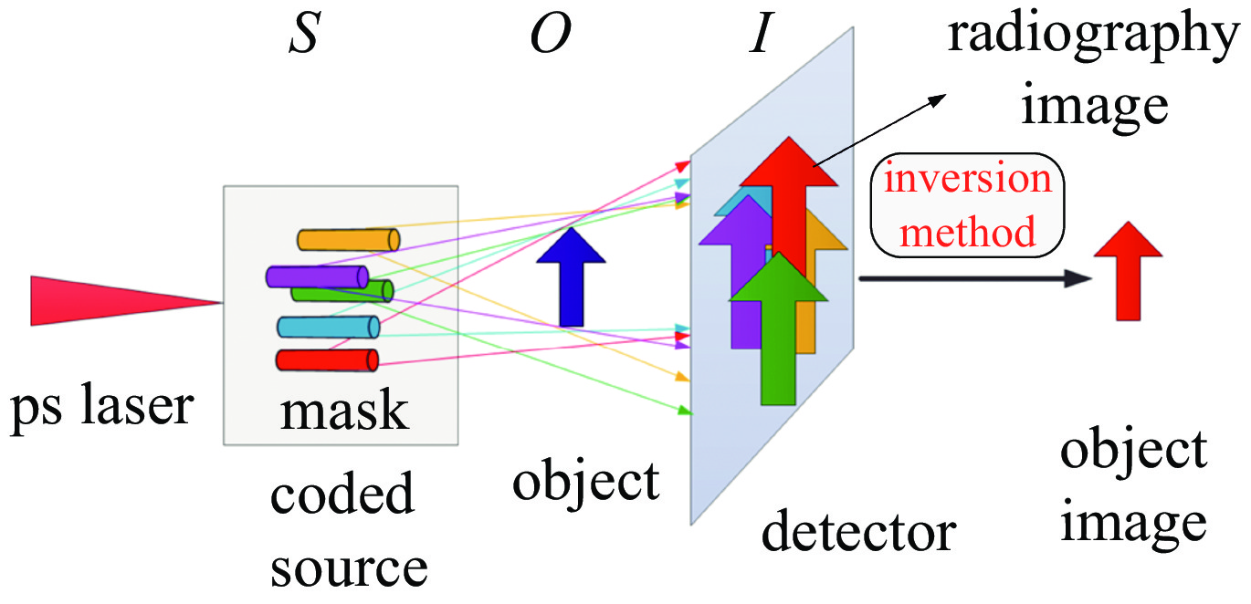
    Fig. 1. Diagram of laser driven X-ray source coding radiography
    Simulated image using pinhole-array plate
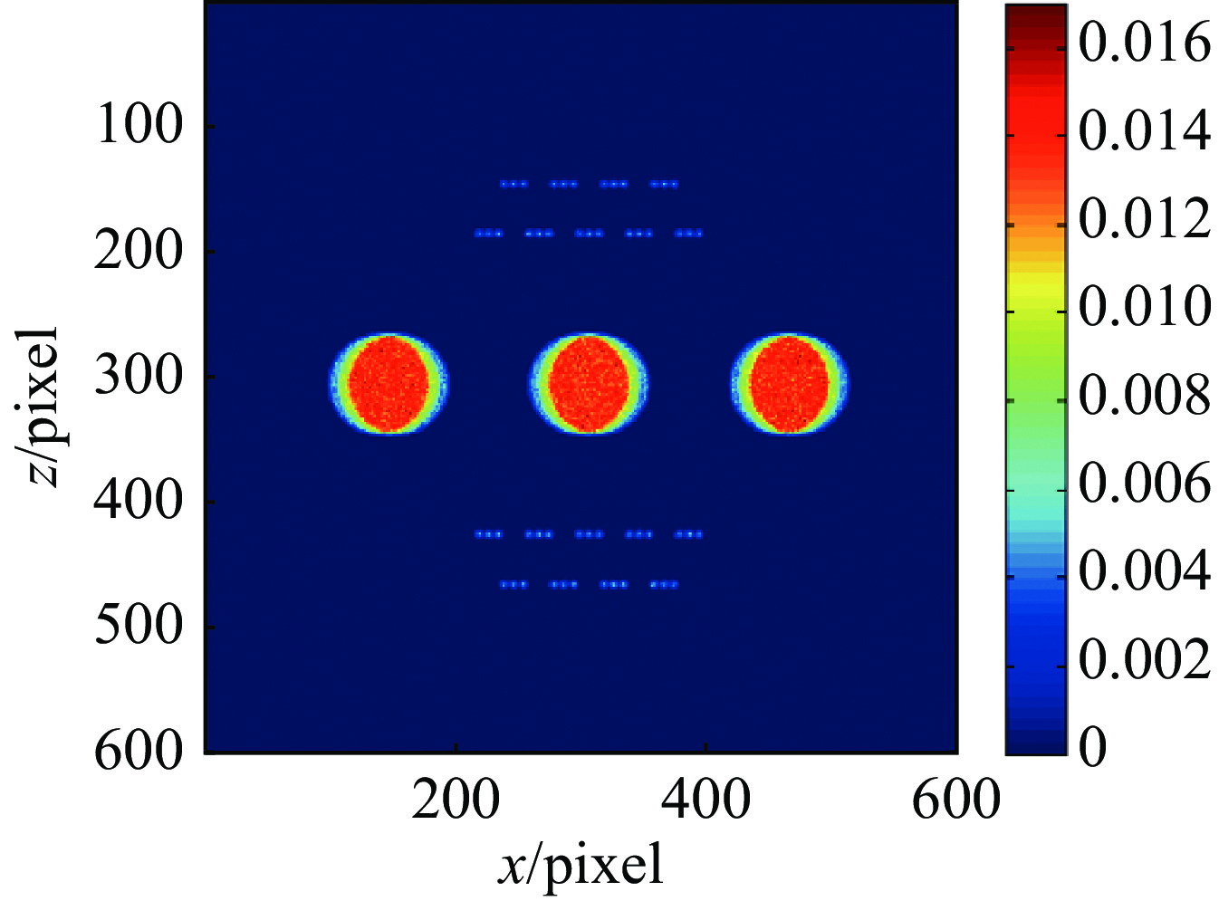
    Fig. 2. Simulated image using pinhole-array plate
    Simulated images by pinhole with different diameter
    Fig. 3. Simulated images by pinhole with different diameter
    Simulated penumbral image and corresponding reconstructed source image at different X-ray brightness
    Fig. 4. Simulated penumbral image and corresponding reconstructed source image at different X-ray brightness
    Radiography image by single-wire target with different X-ray spectrum
    Fig. 5. Radiography image by single-wire target with different X-ray spectrum
    Inversion image from simulated image by wire-array target with different X-ray spectrum
    Fig. 6. Inversion image from simulated image by wire-array target with different X-ray spectrum
    Experimental configuration
    Fig. 7. Experimental configuration
    Design drawing of object and experimental radiography image
    Fig. 8. Design drawing of object and experimental radiography image
    Micrograph picture of assembled wire-array target
    Fig. 9. Micrograph picture of assembled wire-array target
    Pinhole image, penumbral image and reconstructed image from penumbral image of wire-array target
    Fig. 10. Pinhole image, penumbral image and reconstructed image from penumbral image of wire-array target
    Radiography image with wire-array target, inversion image and radiography image with single-wire target
    Fig. 11. Radiography image with wire-array target, inversion image and radiography image with single-wire target
    Edge spread curves from inversion image by wire-array target and radiography image by single-wire target
    Fig. 12. Edge spread curves from inversion image by wire-array target and radiography image by single-wire target
    Bremsstrahlung spectrum of single-wire target and wire-array target
    Fig. 13. Bremsstrahlung spectrum of single-wire target and wire-array target
    Tiankui Zhang, Lianqiang Shan, Minghai Yu, Feng Lu, Weimin Zhou, Chao Tian, Fang Tan, Yonghong Yan, Feng Zhang, Zongqiang Yuan, Qiuyue Xu, Weiwu Wang, Zhigang Deng, Jian Teng, Dongxiao Liu, Lei Yang, Wei Fan, Yue Yang, Kainan Zhou, Jingqin Su, Yuchi Wu, Yongkun Ding, Yuqiu Gu. Source-coded radiography technique with high spatial-resolution for X-ray source driven by ps-laser[J]. High Power Laser and Particle Beams, 2022, 34(12): 122001
    Download Citation