
Ying-ying LI, Zhi-qing ZHANG, Xiao-hong WU, Hsitien Shen Andy. Photoluminescence in Indonesian Fossil Resins[J]. Spectroscopy and Spectral Analysis, 2022, 42(3): 814
- Spectroscopy and Spectral Analysis
- Vol. 42, Issue 3, 814 (2022)
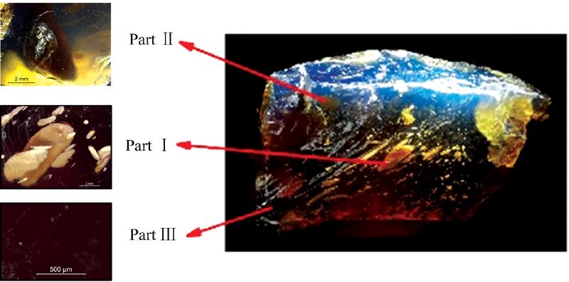
Fig. 1. The blue appearance of IN-5 when under a strong white light. It was divided into three parts: white inclusions (Part Ⅰ), dark inclusions (Part Ⅱ) and basal body (Part Ⅲ)
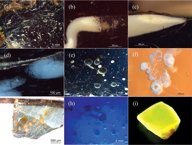
Fig. 2. Images of the inclusion details
(a): The fluid-like white inclusions; (b), (c): The crumb-like white inclusions; (d): The flat-shaped inclusions with spongiform structures; (e): The fried-egg-like dark inclusions with white rims; (f): Cavity-like white inclusion with flower-like structure; (g): The isolated dark and white inclusions in the basal body; (h): Fluorescence of Part Ⅰ—Ⅲ in IN-3 under a 365 nm ultraviolet light; (i): Bright greenish-yellow phosphorescence from IN-4 fragment excited by a 365 nm ultraviolet light
(a): The fluid-like white inclusions; (b), (c): The crumb-like white inclusions; (d): The flat-shaped inclusions with spongiform structures; (e): The fried-egg-like dark inclusions with white rims; (f): Cavity-like white inclusion with flower-like structure; (g): The isolated dark and white inclusions in the basal body; (h): Fluorescence of Part Ⅰ—Ⅲ in IN-3 under a 365 nm ultraviolet light; (i): Bright greenish-yellow phosphorescence from IN-4 fragment excited by a 365 nm ultraviolet light
Fig. 3. FTIR spectra of IN-3, IN-5, and IN-6
Fig. 4. 3D fluorescent patterns of three parts from IN-5 sample
[(a): Part Ⅰ; (b): Part Ⅱ; (c): Part Ⅲ] and bar graph of the ratio of the emission intensity at 446 and 388 nm (d)
[(a): Part Ⅰ; (b): Part Ⅱ; (c): Part Ⅲ] and bar graph of the ratio of the emission intensity at 446 and 388 nm (d)
Fig. 5. Phosphorescence curves and the full width at half maximum (FWHM) in three parts. (a) IN-1 emits stronger phosphorescence by the duration of 365 nm excitation increasing; (b)—(f) Phosphorescence curves and the FWHM of three parts in IN-2—IN-6, respectively
|
Table 1. List of the calculated phosphorescence lifetimes of Parts Ⅰ to Ⅲ, and each R -squared (R 2)

Set citation alerts for the article
Please enter your email address
© Copyright 2018-2021 | Chinese Laser Press. All Rights Reserved 沪ICP备15018463号-20