
Chaoqin SHU, Min ZHU, Yufang ZHU. Cobalt-incorporated Chlorapatite: Preparation by Molten Salt Method, Anti-oxidation and Cytocompatibility [J]. Journal of Inorganic Materials, 2022, 37(11): 1225
- Journal of Inorganic Materials
- Vol. 37, Issue 11, 1225 (2022)
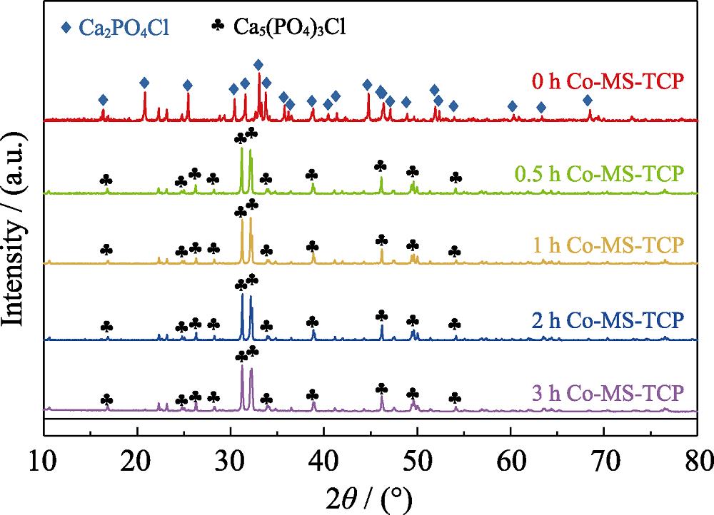
1. XRD patterns of Co-MS-TCP powders prepared by molten salt method with different holding time
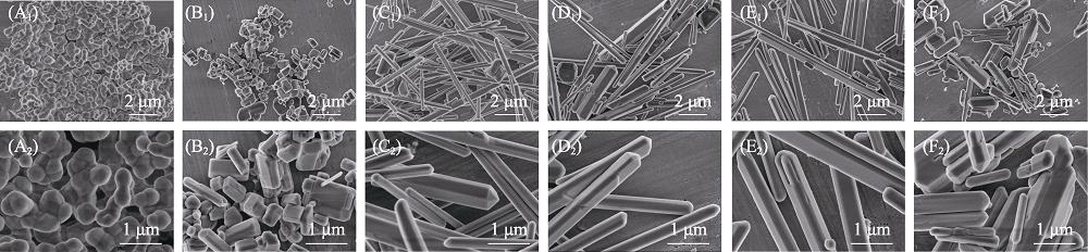
2. SEM images of β -TCP powders before and after molten salt treatment with different holding time
3. H2O2 scavenging effect of (A) MS-TCP and (B) Co-MS-TCP powders prepared by molten salt method with different holding time
4. XRD patterns of Co-MS-TCP powders prepared by molten salt method with different cobalt salt additions
5. SEM images of Co-MS-TCP powders prepared by molten salt method with different cobalt salt additions
6. Contents and valence state of cobalt in Co-MS-TCP on H2O2 scavenging ability
7. Releases of (A) Ca, (B) P and (C) Co ions from β -TCP, MS-TCP and 3% Co-MS-TCP powders in Tris-HCl buffer
8. SEM images of (A1, A2) β -TCP, (B1, B2) MS-TCP and (C1, C2) 3% Co-MS-TCP after soaking in SBF for 7 d and (D) their corresponding FT-IR spectra
9. Cell viabilities of MS-TCP and 3% Co-MS-TCP powders with different concentrations on (A) cartilage cells and (B) bone marrow mesenchymal stem cells
10. Images of intracellular ROS staining fluorescence in cartilage cells stimulated by H2O2 treatment with powders (A) Blank, (B) β -TCP, (C) MS-TCP, (D) 3% Co-MS-TCP and (E) corresponding fluorescence intensity statistics
11. Images of intracellular ROS staining fluorescence in bone marrow mesenchymal stem cells stimulated by H2O2 treatment with powders (A) Blank, (B) β -TCP, (C) MS-TCP, (D) 3% Co-MS-TCP and (E) Corresponding fluorescence intensity statistics.
12. Calcein AM and PI fluorescence images of cartilage cells treated with different powders((A) Blank; (B) β -TCP; (C) MS-TCP; (D) 3% Co-MS-TCP; (A1-D1) Cells without H2O2 stimulation; (A2-D2) Cells stimulated with H2O2 showing live (green) and death (red) cells)
13. Calcein AM and PI fluorescence images of bone marrow mesenchymal stem cells treated with powders (A) Blank; (B) β -TCP; (C) MS-TCP; (D) 3% Co-MS-TCP; (A1-D1) Cells without H2O2 stimulation; (A2-D2) Cells stimulated with H2O2 showing live (green) and death (red) cells


Set citation alerts for the article
Please enter your email address

© Copyright 2018-2021 | Chinese Laser Press. All Rights Reserved 沪ICP备15018463号-20