• Laser & Optoelectronics Progress
  • Vol. 60, Issue 1, 0129001 (2023)
Wenquan Qiu, Wei Liu*, Hongyan Jia, Tiantian Qi, Jin Shen, and Yajing Wang
Author Affiliations
  • School of Electrical and Electronic Engineering, Shandong University of Technology, Zibo 255049, Shandong , China
  • show less
    DOI: 10.3788/LOP212992 Cite this Article Set citation alerts
    Wenquan Qiu, Wei Liu, Hongyan Jia, Tiantian Qi, Jin Shen, Yajing Wang. Influence of Different Structures of Capillary Cell on Electric Field Intensity in Detection Area[J]. Laser & Optoelectronics Progress, 2023, 60(1): 0129001 Copy Citation Text show less
    Schematic diagram of the optical path and detection position of the capillary cell
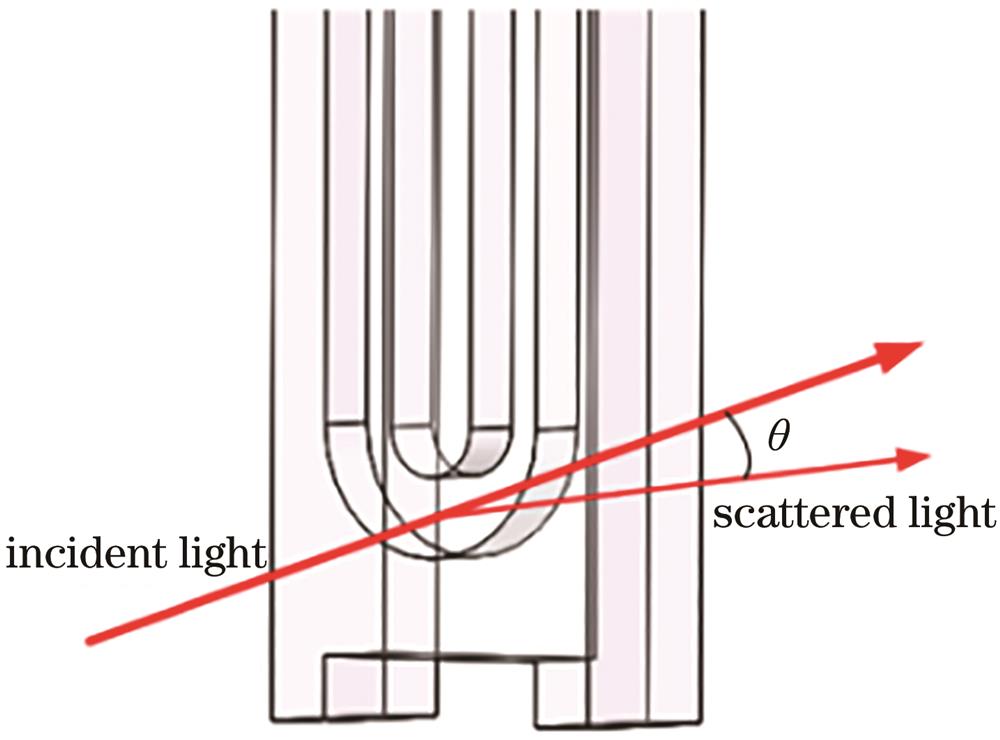
    Fig. 1. Schematic diagram of the optical path and detection position of the capillary cell
    Capillary cell simulation model
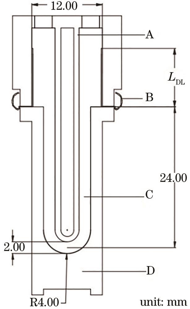
    Fig. 2. Capillary cell simulation model
    Schematic diagram of the detection point of the capillary cell
    Fig. 3. Schematic diagram of the detection point of the capillary cell
    Influence of electrode length on the electric field intensity. (a) Influence of electrode length on the electric field intensity of different detection positions; (b) influence of different electrode lengths on the electric field intensity of the detection center
    Fig. 4. Influence of electrode length on the electric field intensity. (a) Influence of electrode length on the electric field intensity of different detection positions; (b) influence of different electrode lengths on the electric field intensity of the detection center
    Relationship between electrode length and electric field intensity change rate
    Fig. 5. Relationship between electrode length and electric field intensity change rate
    Models of capillary cells with different structures
    Fig. 6. Models of capillary cells with different structures
    Electric field intensity of the semicircular capillary cell. (a) Overall electric field intensity diagram; (b) electric field intensity in the detection area
    Fig. 7. Electric field intensity of the semicircular capillary cell. (a) Overall electric field intensity diagram; (b) electric field intensity in the detection area
    Electric field intensity of the U-shaped capillary cell. (a) Overall electric field intensity diagram; (b) electric field intensity in the detection area
    Fig. 8. Electric field intensity of the U-shaped capillary cell. (a) Overall electric field intensity diagram; (b) electric field intensity in the detection area
    Electric field intensity of the inverted Ω-shaped capillary cell. (a) Overall electric field intensity diagram; (b) electric field intensity in the detection area
    Fig. 9. Electric field intensity of the inverted Ω-shaped capillary cell. (a) Overall electric field intensity diagram; (b) electric field intensity in the detection area
    Electric field intensity of the H-shaped capillary cell. (a) Overall electric field intensity diagram; (b) electric field intensity in the detection area
    Fig. 10. Electric field intensity of the H-shaped capillary cell. (a) Overall electric field intensity diagram; (b) electric field intensity in the detection area
    Electric field intensity of the triangular capillary cell. (a) Overall electric field intensity diagram; (b) electric field intensity in the detection area
    Fig. 11. Electric field intensity of the triangular capillary cell. (a) Overall electric field intensity diagram; (b) electric field intensity in the detection area
    Changes of electric field intensity in the detection area of capillary cells with different structures
    Fig. 12. Changes of electric field intensity in the detection area of capillary cells with different structures
    ComponentMaterialRelative permittivityConductivity /(S·m-1
    ElectrodeGold-plated copper1.04.10×107
    Capillary cell wallPolycarbonate3.23.15×10-13
    Table 1. Capillary cell material properties
    Sample cell typeElectric field intensity /(V·m-1
    n=0.6 mmn=0.4 mmn=0.2 mmn=0 mmn=-0.2 mmn=-0.4 mmn=-0.6 mm
    Semicircular capillary cell3270301628012615245123092190
    U-shaped capillary cell2787275327132670262925932566
    Inverted Ω-shaped cell2605259625862575256425542547
    H-shaped capillary cell2735271526912665264026192603
    Triangular capillary cell2582257825732567255825512546
    Table 2. Electric field intensity values at different positions in the detection area of the capillary cell
    Sample cell typeElectric field intensity change rate /%
    n=0.6 mmn=0.4 mmn=0.2 mmn=0 mmn=-0.2 mmn=-0.4 mmn=-0.6 mm
    Semicircular capillary cell25.0515.337.110.00-6.27-11.70-16.25
    U-shaped capillary cell4.383.111.610.00-1.54-2.88-3.90
    Inverted Ω-shaped cell1.170.820.430.00-0.43-0.82-1.09
    H-shaped capillary cell2.631.880.980.00-0.94-1.73-2.33
    Triangular capillary cell0.580.430.230.00-0.35-0.62-0.82
    Table 3. Electric field intensity change rate at different positions in the detection area of the capillary cell
    Sample cell typeBending coefficientElectric field intensity at the detection center /(V·m-1Electric field intensity change rate /%
    Semicircular capillary cell1.00261533.02
    U-shaped capillary cell0.8826707.93
    H-shaped capillary cell0.6926654.83
    Inverted Ω-shaped cell0.5725752.23
    Triangular capillary cell0.2225671.39
    Table 4. Influence of the bending degree of the capillary cell on the electric field intensity at the detection center
    Sample cell typeAxial distance between the electrodes /mmElectric field intensity at the detection center /(V·m-1Average value of electric field intensity /(V·m-1
    Semicircular capillary cell67.1326152652
    U-shaped capillary cell68.4026702673
    H-shaped capillary cell69.7026652666
    Inverted Ω-shaped cell69.8825752575
    Triangular capillary cell70.6825672565
    Table 5. Relationship of the axial distance between electrodes and electric field intensity at the detection center
    Wenquan Qiu, Wei Liu, Hongyan Jia, Tiantian Qi, Jin Shen, Yajing Wang. Influence of Different Structures of Capillary Cell on Electric Field Intensity in Detection Area[J]. Laser & Optoelectronics Progress, 2023, 60(1): 0129001
    Download Citation