Cancer is a disease caused by the uncontrolled growth and division of malignant cancer cells. Since the 21st century, the incidence and mortality of cancer have been increasing rapidly worldwide, making it a medical problem that affects countries worldwide. The pathological mechanism of cancer and the study of various therapeutics based on inducing cancer cell death both greatly benefit from research on the morphology and function of cancer cells at the single-cell level, particularly research on the process of cancer cell death. Digital holographic microscopy, a quantitative phase imaging technique, offers a nondestructive, unlabeled, and noncontact quantitative measurement tool for biological research. It can also provide nondestructive quantitative imaging of living cells. In this paper, digital holographic tomography was used in the three-dimensional quantitative detection of bladder cancer cell vacuolation. This work can broaden the field of application for digital holographic tomography in the biomedical industry, offer new perspectives on how to study the morphological changes that occur during cancer cell apoptosis, and investigate potential new cancer treatment approaches.
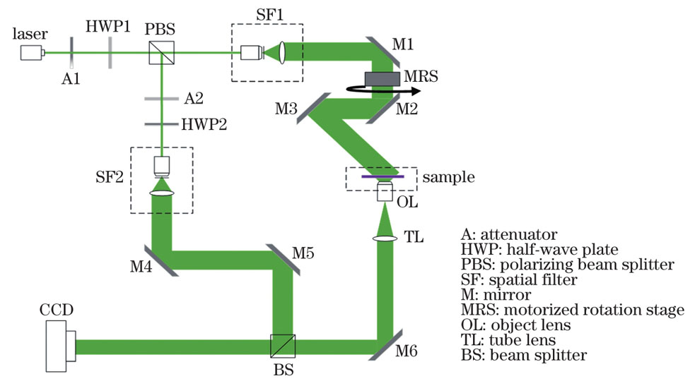
This study focuses on the vacuolar structure of cancer cells existing in the process of paraptosis. First, the hologram of bladder cancer cells with vacuoles inside was obtained using digital holographic microscopy. Then, the amplitude and phase of cells were obtained by filtering, digital focusing, angular spectrum propagation, and phase unwrapping. Their three-dimensional morphology and spatial locations were reconstructed using the diffraction tomography reconstruction algorithm combined with nonnegative constraints. Finally, morphological parameters such as the surface and volume of the vacuoles were calculated according to the number of pixels in the image.
Digital holographic tomography was used in this study to produce three-dimensional reconstruction results of four bladder cancer cells with vacuoles (Figure 8). Four morphological parameters, including the volume, surface, surface to volume, and the ratio of vacuoles volume to cell volume, were calculated (Table 1). Digital holographic tomography, as a technique for three-dimensional quantitative imaging, was used to examine cancer cells with vacuoles. It can quantitatively determine the volume, position, and other morphological parameters of the vacuoles. By combining with the biomedical research, it can be used to observe the changes in three-dimensional shape and volume of tumor cells’ internal vacuoles induced by drugs, to explore the correlation between the expression of some proteins and the morphological characteristics of vacuoles, to provide a more comprehensive and profound understanding of the process of paraptosis of cancer cells and to find new methods for cancer treatment.
The application of digital holographic tomography to bladder cancer cell vacuolation imaging is described in this research. The results have shown that digital holographic tomography can accurately reconstruct the three-dimensional shape and space position of the vacuolation in bladder cancer cells. The above research progress is of great significance for studying the paraptosis process of cancer cells as well as the related mechanisms and treatment strategies of drug-induced paraptosis of cancer cells.