• Acta Physica Sinica
  • Vol. 69, Issue 16, 168202-1 (2020)
Qiong Wang1、2, Kai-Ge Wang1、*, Kang Kang Meng1, Dan Sun1, Tong Yu Han1, and Ai-Hua Gao2、*
Author Affiliations
  • 1National Center for International Research of Photoelectric Technology and Nano-Functional Materials and Application, State Key Laboratory of Cultivation Base for Photoelectric Technology and Functional Materials, Laboratory of Photoelectric Technology of Shaanxi Province, Institute of Photonics and Photon-Technology, Northwest University, Xi’an 710069, China
  • 2School of Physics, Northwestern University, Xi’an 710069, China
  • show less
    DOI: 10.7498/aps.69.20200074 Cite this Article
    Qiong Wang, Kai-Ge Wang, Kang Kang Meng, Dan Sun, Tong Yu Han, Ai-Hua Gao. Electrodynamic characteristics of λ-DNA molecule translocating through the microfluidic channel port studied with single molecular fluorescence imaging technology[J]. Acta Physica Sinica, 2020, 69(16): 168202-1 Copy Citation Text show less
    Schematic diagram of experimental set-up.
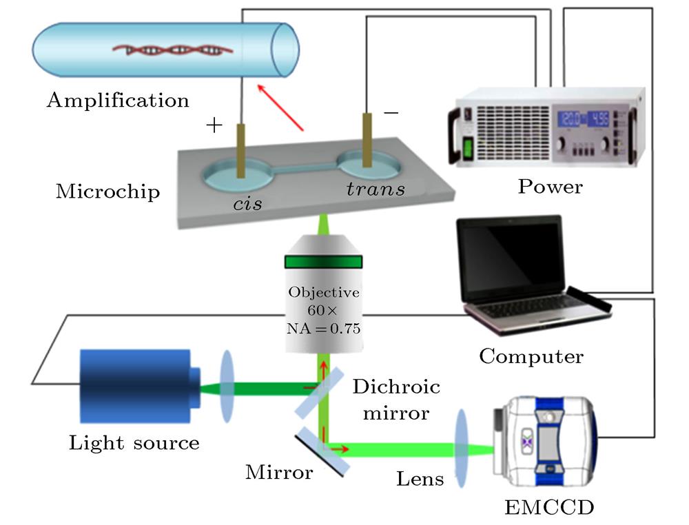
    Fig. 1. Schematic diagram of experimental set-up.
    DNA molecules enter the microchannel from the trans port and migrate inside (E = 3.75 × 10³ V·m–1): (a) CCD photographs; (b) DNA molecular position.
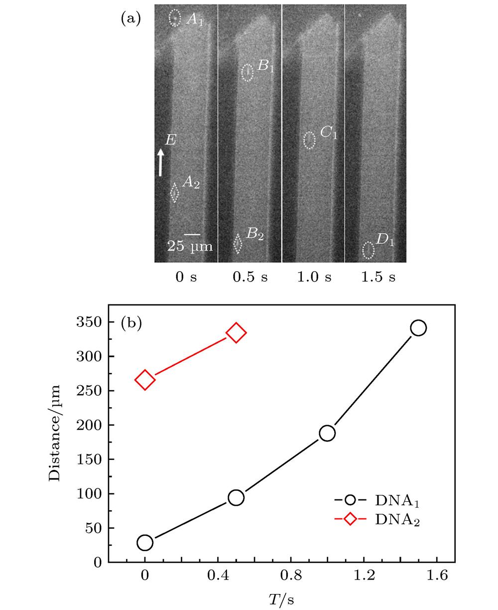
    Fig. 2. DNA molecules enter the microchannel from the trans port and migrate inside (E = 3.75 × 10³ V·m–1): (a) CCD photographs; (b) DNA molecular position.
    The velocity of DNA molecules entering and leaving the port (E = 3.75 × 10³ V·m–1): (a) Entering the trans port; (b) leaving the cis port; and (c) velocity versus electric intensity.
    Fig. 3. The velocity of DNA molecules entering and leaving the port (E = 3.75 × 10³ V·m–1): (a) Entering the trans port; (b) leaving the cis port; and (c) velocity versus electric intensity.
    Reversed motion of DNA molecules within micro channel under different electric intensity: (a) E = 8.125 × 10³ V·m–1; (b) E = 9.375 × 10³ V·m–1; (c) percentage of DNA molecules with reversal motion direction in different regions of the cis port under different electric intensity.
    Fig. 4. Reversed motion of DNA molecules within micro channel under different electric intensity: (a) E = 8.125 × 10³ V·m–1; (b) E = 9.375 × 10³ V·m–1; (c) percentage of DNA molecules with reversal motion direction in different regions of the cis port under different electric intensity.
    The motion of DNA molecules near the trans port: (a) Reciprocating along the axis; (b) rotating.
    Fig. 5. The motion of DNA molecules near the trans port: (a) Reciprocating along the axis; (b) rotating.
    The track of DNA molecules reciprocating near the trans port (E = 9.375 × 10³ V·m–1).
    Fig. 6. The track of DNA molecules reciprocating near the trans port (E = 9.375 × 10³ V·m–1).
    Aggregates of DNA molecules on the wall of microchannel near the trans port; (a) E = 7.5 × 10³ V·m–1; (b) E = 1 × 10⁴ V·m–1.
    Fig. 7. Aggregates of DNA molecules on the wall of microchannel near the trans port; (a) E = 7.5 × 10³ V·m–1; (b) E = 1 × 10⁴ V·m–1.
    Schematic diagram of buffer velocity distribution in microfluidic channel and the infromation of DNA: (a) Force; (b) velocity.
    Fig. 8. Schematic diagram of buffer velocity distribution in microfluidic channel and the infromation of DNA: (a) Force; (b) velocity.
    Measured and theoretical velocities of DNA molecules at different positions on the same cross section of near the microchannel port.
    Fig. 9. Measured and theoretical velocities of DNA molecules at different positions on the same cross section of near the microchannel port.
    Schematic diagram of DNA molecules moving near the port of microchannel: (a) reversing near the cis port, and the reversed DNA molecule is easy to be adsorbed onto the inner wall, 7.5 × 10³ V·m–1 ≤ E ≤ 1 × 10⁴ V·m–1; (b) reversing near the trans port, E > 1 × 10 4 V·m–1.
    Fig. 10. Schematic diagram of DNA molecules moving near the port of microchannel: (a) reversing near the cis port, and the reversed DNA molecule is easy to be adsorbed onto the inner wall, 7.5 × 10³ V·m–1E ≤ 1 × 10⁴ V·m–1; (b) reversing near the trans port, E > 1 × 10 4 V·m–1.
    Qiong Wang, Kai-Ge Wang, Kang Kang Meng, Dan Sun, Tong Yu Han, Ai-Hua Gao. Electrodynamic characteristics of λ-DNA molecule translocating through the microfluidic channel port studied with single molecular fluorescence imaging technology[J]. Acta Physica Sinica, 2020, 69(16): 168202-1
    Download Citation