• Chinese Optics Letters
  • Vol. 13, Issue 7, 071002 (2015)
Jiulou Zhang1, Junwei Shi1, Simin Zuo1, Fei Liu1、2, Jing Bai1, and Jianwen Luo1、3、*
Author Affiliations
  • 1Department of Biomedical Engineering, School of Medicine, Tsinghua University, Beijing 100084, China
  • 2Tsinghua-Peking Center for Life Sciences, Beijing 100084, China
  • 3Center for Biomedical Imaging Research, Tsinghua University, Beijing 100084, China
  • show less
    DOI: 10.3788/COL201513.071002 Cite this Article Set citation alerts
    Jiulou Zhang, Junwei Shi, Simin Zuo, Fei Liu, Jing Bai, Jianwen Luo. Fast reconstruction in fluorescence molecular tomography using data compression of intra- and inter-projections[J]. Chinese Optics Letters, 2015, 13(7): 071002 Copy Citation Text show less
    The results of the phantom experiment. (a) Slice and (g) 3D view of the true double fluorescence targets. (b) Slice and (h) 3D view of the reconstructed images obtained from the conventional method without data compression. (c)–(f) Slice and (i)–(l) 3D view of the reconstructed images obtained from the FDC strategy with 2, 3, 4, and 6 adjacent projections in each group. (m) Computational time consumed using different methods. (n) RMSEs and (o) normalized intensity profiles along the dotted line in (a) using different methods. All images are normalized by the maximal values of the results.
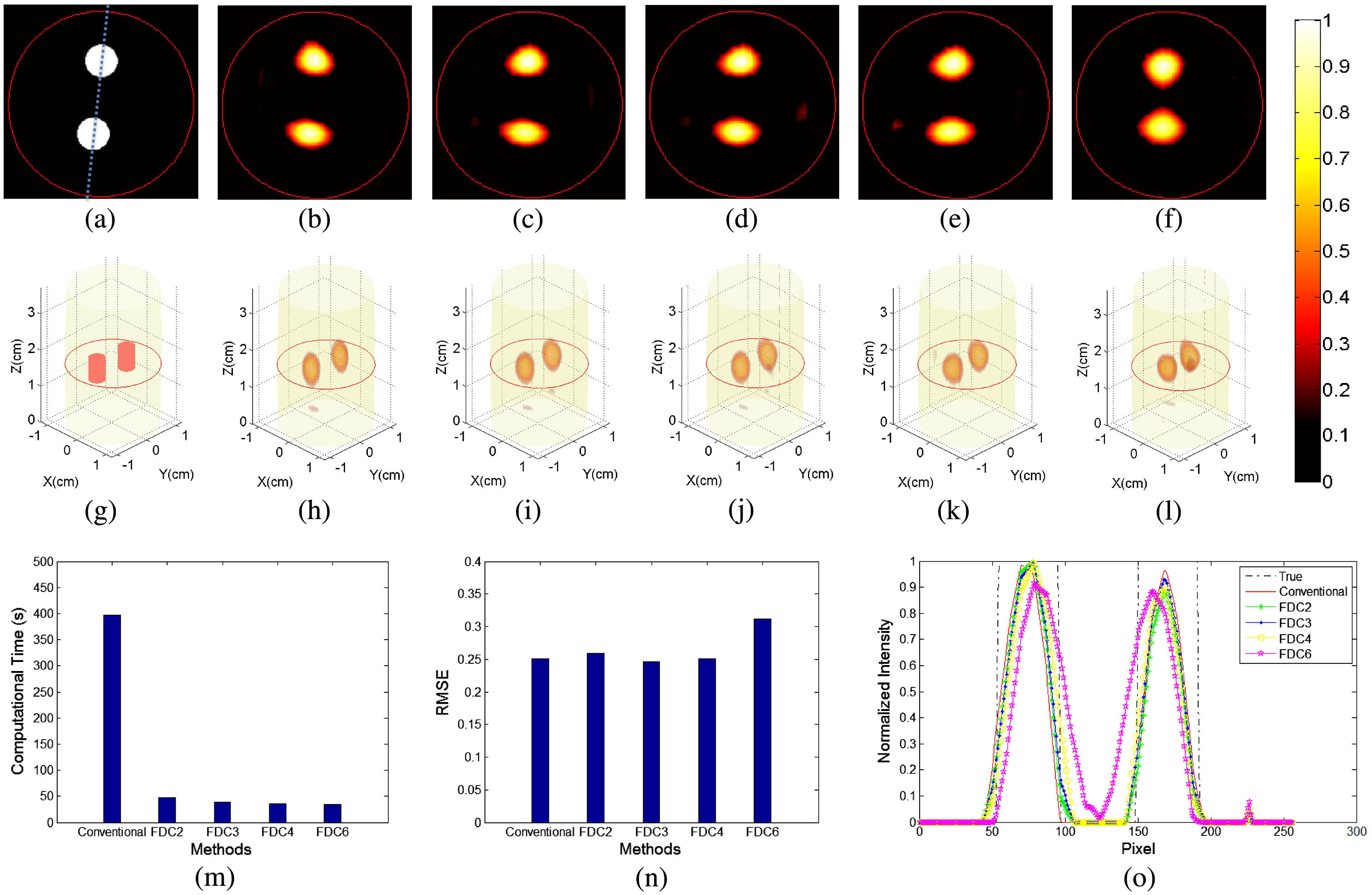
    Fig. 1. The results of the phantom experiment. (a) Slice and (g) 3D view of the true double fluorescence targets. (b) Slice and (h) 3D view of the reconstructed images obtained from the conventional method without data compression. (c)–(f) Slice and (i)–(l) 3D view of the reconstructed images obtained from the FDC strategy with 2, 3, 4, and 6 adjacent projections in each group. (m) Computational time consumed using different methods. (n) RMSEs and (o) normalized intensity profiles along the dotted line in (a) using different methods. All images are normalized by the maximal values of the results.
    Results of the in vivo mouse experiment. (a) Slice view of the in vivo mouse. (b) Slice and (f) 3D view of the reconstructed images obtained from the conventional method. (c)–(e) Slice and (g)–(i) 3D view of the reconstructed images obtained from the FDC with 2, 3, and 6 adjacent projections. (j) Computational time consumed using different methods. (k) RMSEs and (l) normalized intensity profiles along the dotted line in (a) using different methods. All images are normalized by the maximal values of the results.
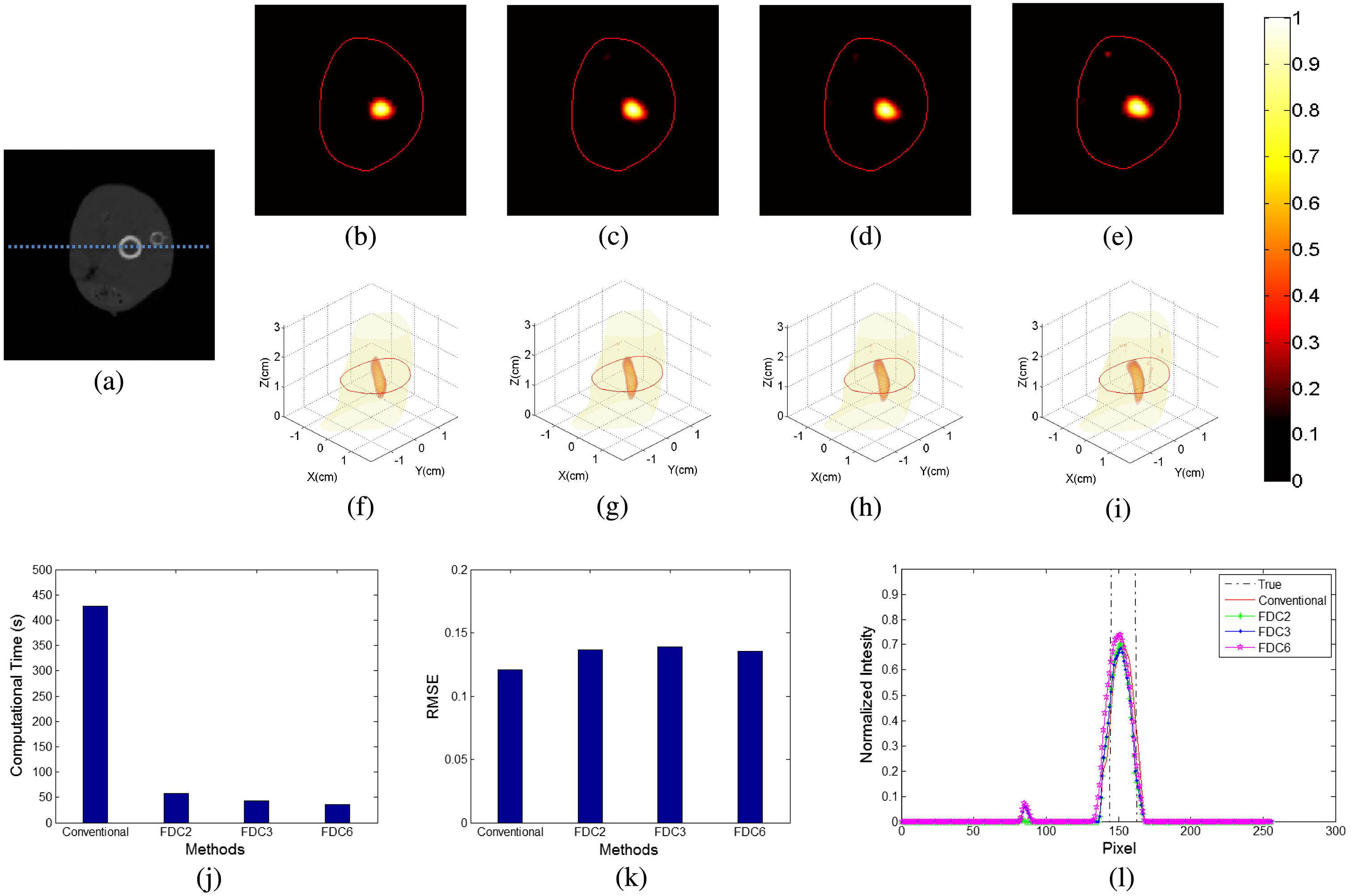
    Fig. 2. Results of the in vivo mouse experiment. (a) Slice view of the in vivo mouse. (b) Slice and (f) 3D view of the reconstructed images obtained from the conventional method. (c)–(e) Slice and (g)–(i) 3D view of the reconstructed images obtained from the FDC with 2, 3, and 6 adjacent projections. (j) Computational time consumed using different methods. (k) RMSEs and (l) normalized intensity profiles along the dotted line in (a) using different methods. All images are normalized by the maximal values of the results.
    MethodsPhantomIn vivo
    Conventional13764×1502813576×15231
    FDC23933×150284150×15231
    FDC32888×150282875×15231
    FDC42223×15028Null
    FDC61558×150281500×15231
    Table 1. The Scales of Weight Matrix before and after Compression
    Jiulou Zhang, Junwei Shi, Simin Zuo, Fei Liu, Jing Bai, Jianwen Luo. Fast reconstruction in fluorescence molecular tomography using data compression of intra- and inter-projections[J]. Chinese Optics Letters, 2015, 13(7): 071002
    Download Citation