• Laser & Optoelectronics Progress
  • Vol. 57, Issue 2, 21003 (2020)
Qiu Yue1, Tang Chen1、*, Xu Min1, Huang Shengjian1, and Lei Zhenkun2
Author Affiliations
  • 1School of Electrical and Information Engineering, Tianjin University, Tianjin 300072, China
  • 2State Key Laboratory of Structural Analysis for Industrial Equipment, Dalian University of Technology, Dalian, Liaoning 116023, China
  • show less
    DOI: 10.3788/LOP57.021003 Cite this Article Set citation alerts
    Qiu Yue, Tang Chen, Xu Min, Huang Shengjian, Lei Zhenkun. Shearlet-Transform-Based Improved Total Variation Speckle Denoising Method[J]. Laser & Optoelectronics Progress, 2020, 57(2): 21003 Copy Citation Text show less
    Flow chart of algorithm
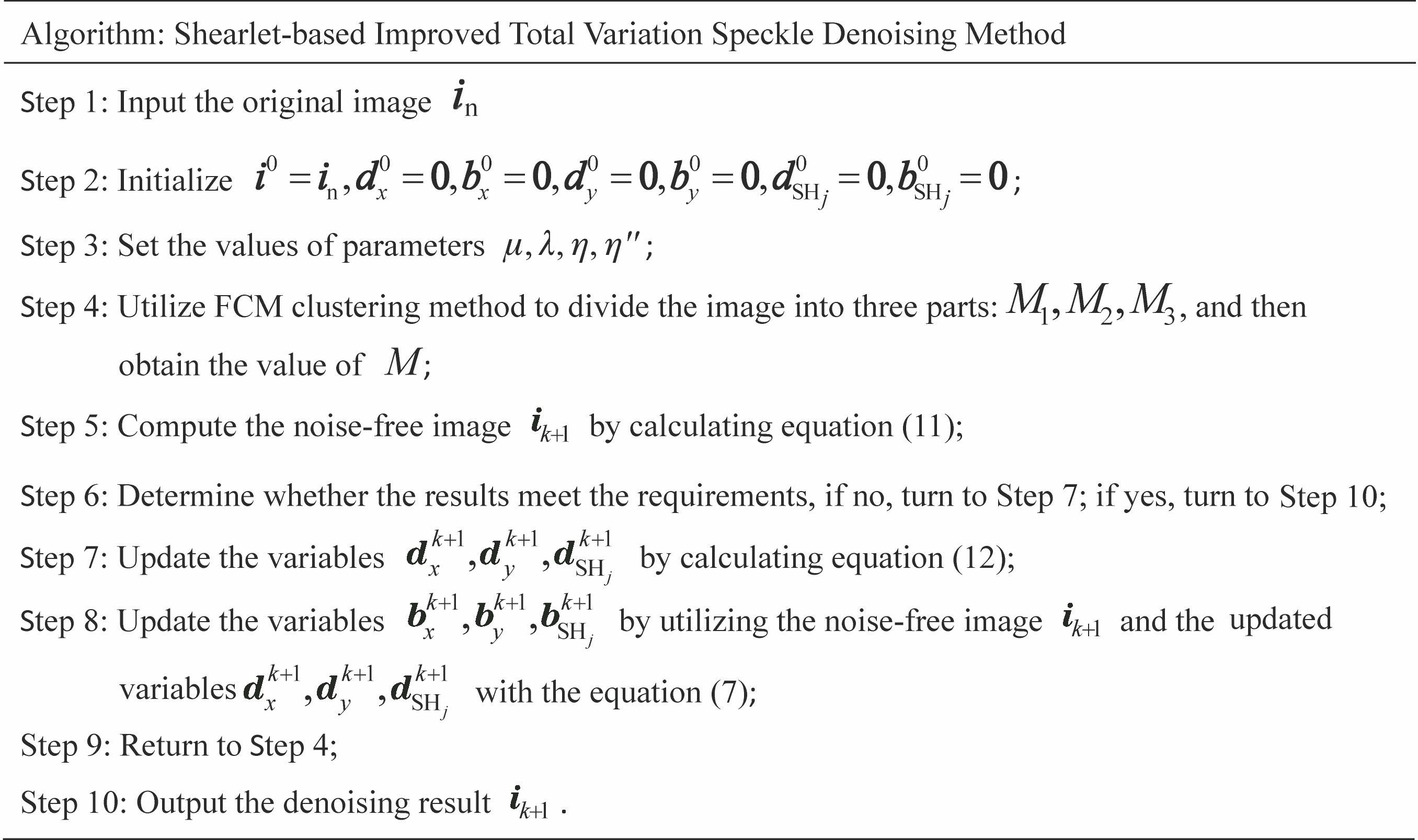
    Fig. 1. Flow chart of algorithm
    OCT B-scan images of human retina. (a) Normal retina image; (b) DME retina image; (c) Drusen retina image
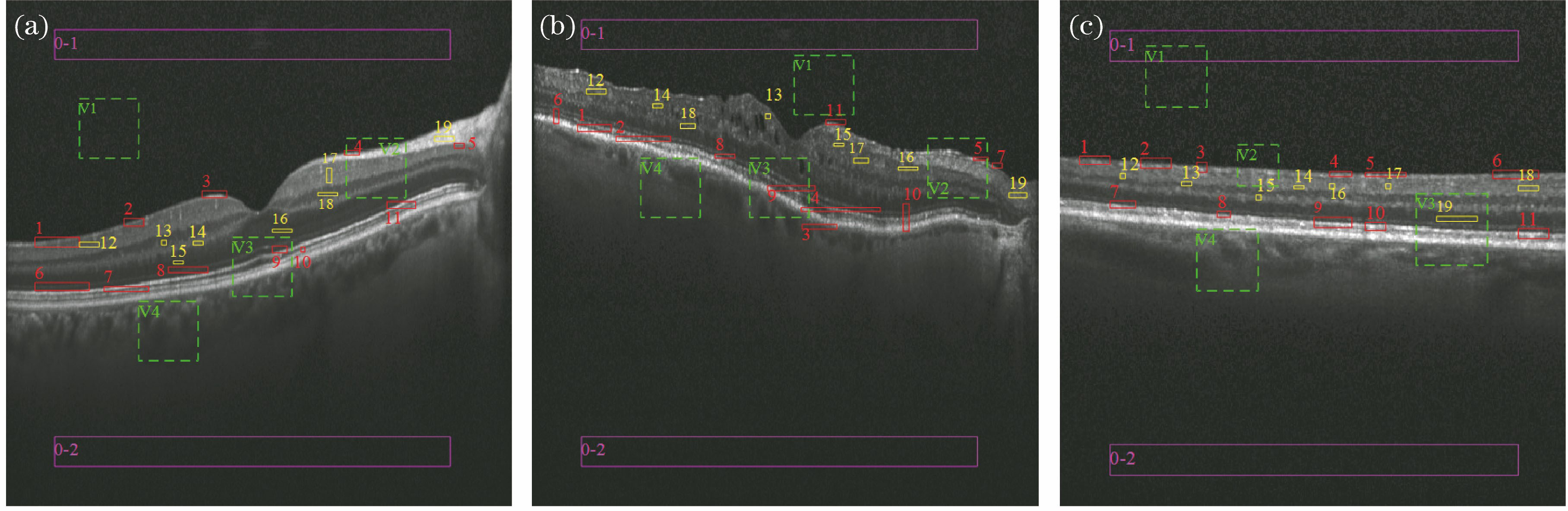
    Fig. 2. OCT B-scan images of human retina. (a) Normal retina image; (b) DME retina image; (c) Drusen retina image
    Speckle denoising of normal retina image. (a0)--(a4) Original OCT image with noise and images after speckle denoising by ACD, Curvelet, TV, and proposed method; (b0)--(b4) histograms corresponding to images in Figs. 3 (a0)--(a4); (c10)--(c44) enlarged ROIs V1--V4 corresponding to images in Figs. 3(a0)--(a4)
    Fig. 3. Speckle denoising of normal retina image. (a0)--(a4) Original OCT image with noise and images after speckle denoising by ACD, Curvelet, TV, and proposed method; (b0)--(b4) histograms corresponding to images in Figs. 3 (a0)--(a4); (c10)--(c44) enlarged ROIs V1--V4 corresponding to images in Figs. 3(a0)--(a4)
    Speckle denoising of DME retina image. (a0)--(a4) Original OCT image with noise and images after speckle denoising by ACD, Curvelet, TV, and proposed method; (b0)--(b4) histograms corresponding to images in Figs. 4 (a0)--(a4); (c10)--(c44) enlarged ROIs V1--V4 corresponding to images in Figs. 4(a0)--(a4)
    Fig. 4. Speckle denoising of DME retina image. (a0)--(a4) Original OCT image with noise and images after speckle denoising by ACD, Curvelet, TV, and proposed method; (b0)--(b4) histograms corresponding to images in Figs. 4 (a0)--(a4); (c10)--(c44) enlarged ROIs V1--V4 corresponding to images in Figs. 4(a0)--(a4)
    Speckle denoising of Drusen retina image. (a0)--(a4) Original OCT image and images after speckle denoising by ACD, Curvelet, TV, and proposed method; (b0)--(b4) histograms corresponding to images in Figs. 5(a0)--(a4); (c10)--(c44) enlarged ROIs V1--V4 corresponding to images in Figs. 5(a0)--(a4)
    Fig. 5. Speckle denoising of Drusen retina image. (a0)--(a4) Original OCT image and images after speckle denoising by ACD, Curvelet, TV, and proposed method; (b0)--(b4) histograms corresponding to images in Figs. 5(a0)--(a4); (c10)--(c44) enlarged ROIs V1--V4 corresponding to images in Figs. 5(a0)--(a4)
    MethodSNR /dBENLCNRγ
    Original30.6061444.40004.1625-
    ACD53.6106280.10005.88350.9832
    Curvelet41.91111055.80008.94450.9976
    TV39.94831182.300011.61040.9866
    Proposed48.94511346.800013.27540.9932
    Table 1. Metrics of normal retina image experiment
    MethodSNR /dBENLCNRγ
    Original29.743384.75222.9804-
    ACD44.1283160.84774.35710.9924
    Curvelet40.7001287.33235.42750.9953
    TV37.9425550.23596.31730.9885
    Proposed46.7891649.10498.66470.9927
    Table 2. Metrics of DME retina image experiment
    MethodSNR /dBENLCNRγ
    Original26.8779201.59033.1580-
    ACD44.5433305.33315.63490.9908
    Curvelet42.5725881.42778.27960.9930
    TV38.2609922.40899.24660.9906
    Proposed43.1048940.01249.41480.9934
    Table 3. Metrics of Drusen retina image experiment
    Qiu Yue, Tang Chen, Xu Min, Huang Shengjian, Lei Zhenkun. Shearlet-Transform-Based Improved Total Variation Speckle Denoising Method[J]. Laser & Optoelectronics Progress, 2020, 57(2): 21003
    Download Citation