Medical imaging is a key tool for life science research, diagnosis and treatment. Traditional medical imaging techniques include magnetic resonance imaging (MRI), computed tomography (CT), positron emission tomography (PET), ultrasonic imaging (US) and optical coherence tomography (OCT), etc. Each of them plays an important role in medical imaging, with each showing its own advantages and limitations. MRI is not only expensive but also has many safety restrictions, which means it is not suitable for the patients with pacemakers or claustrophobic. CT is not suitable for children and pregnant women because of ionizing radiation. PET has a large number of applications in the study of life metabolism and tumor research. However, it requires radioactive element markers and is difficult to be widely used in clinical medical imaging due to its high cost. US has poor specificity and spatial resolution, and lacks fine biological structure imaging capabilities for the early development of the disease. OCT and other optical imaging technologies are limited by the optical diffusion limit, which results in the general imaging depth within about 1 mm.
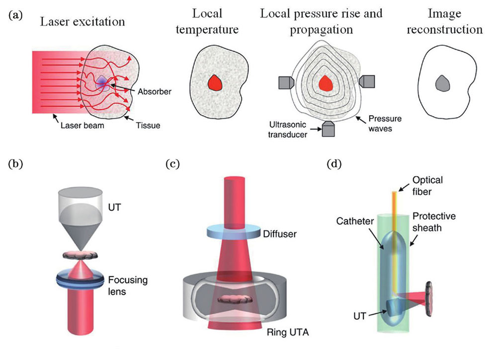
Photoacoustic (PA) imaging is a hybrid biomedical imaging modality combining the advantages of high contrast of optical imaging and deep penetration of ultrasound imaging. The spatial scale of PA imaging covers from subcellular structures to organs. In addition, it has many other advantages such as non-invasive imaging, label-free imaging, molecular imaging and compatible with multi-modality. Although PA imaging has encountered many challenges in the process of clinical translation, PA imaging has overcome a series of difficulties and will have broader application prospects in the field of biomedical imaging thanks to the development of related technologies. The purpose of this article is to help readers in related fields of biomedical imaging to form a more comprehensive understanding of PA imaging, and to quickly understand the main progress of PA imaging research in recent years.
This review article provides a brief introduction to the basic principles and main modes of PA imaging. Photoacoustic computed tomography (PACT) and photoacoustic microscopy imaging (PAM) are the two main modes of PA imaging. Photoacoustic endscopy (PAE) is the application of PA in endoscopy, and photoacoustic molecular imaging expands the capabilities of PA imaging in molecular-level detection.
PACT is suitable for large-scale imaging of the human brain, limbs, breast and other large-size targets. Over the past decade, PACT has made lots of advances in high-speed and deep imaging. However, the issues about economy and portability still hinder the further clinical translation of PACT. Fortunately, the technological development of ultrasonic transducers and low-cost laser sources and the advancement of advanced reconstruction algorithms have provided solutions to the above problems. PAM has broad application prospects from subcellular structure to organ level. High/super-resolution, fast imaging, and higher imaging quality have always been the common pursuit of researchers. The nonlinear effect greatly contributes to the improvement of the resolution of PA imaging. The extended depth-of-field technology can solve the defocus problem encountered by PAM in volume imaging. Advanced scanning methods are one of the main solutions to high-speed PA imaging. In addition, non-contact PA imaging is another important direction in clinical applications, and corresponding technological breakthroughs have also been made in recent years. As the expansion of PA imaging, PAE is a promising technology for endoscopic imaging by drawing on the related progress of PACT and PAM, such as extended depth-of-field technology, optical scanning methods and multi-modality imaging. Aiming at the problem that some tumors lack characteristic absorption peaks, PA molecular imaging holds great promise in the diagnosis and treatment of diseases. In recent years, the development of PA molecular imaging has focused on the near-infrared window to improve the depth of imaging. The PA signal enhancement mechanism can improve the sensitivity and specificity of imaging. Furthermore, the design of integrated diagnosis and treatment PA contrast agents is also a hot topic in PA molecular imaging.
Although PA imaging has made a lot of breakthroughs in recent years, it has not yet achieved a large-scale clinical application. In the next few years, the resolution, depth, speed and sensitivity of PA imaging will still be the research focus of researchers in related fields. Costs will be further reduced by using low-cost laser sources, while imaging speed and quality will meet the needs of most clinical applications thanks to technological advances in related fields. In addition, by the design of the PA contrast agent in the second near-infrared window (NIR-Ⅱ), the depth of PA imaging will further increase. We believe that through the joint efforts of researchers in related fields, PA imaging will play a more important role in the clinic practices.