• Infrared and Laser Engineering
  • Vol. 51, Issue 11, 20220550 (2022)
Jing Yao1、2、3、4, Zhipeng Yu1、2、4, Yufeng Gao3, Shiwei Ye3, Wei Zheng3、*, and Puxiang Lai1、2、4
Author Affiliations
  • 1Department of Biomedical Engineering, the Hong Kong Polytechnic University, Hong Kong SAR, China
  • 2The Hong Kong Polytechnic University Shenzhen Research Institute, Shenzhen 518055, China
  • 3Research Center for Biomedical Optics and Molecular Imaging, Shenzhen Institutes of Advanced Technology, Chinese Academy of Sciences, Shenzhen 518055, China
  • 4Photonics Research Institute, the Hong Kong Polytechnic University, Hong Kong SAR, China
  • show less
    DOI: 10.3788/IRLA20220550 Cite this Article
    Jing Yao, Zhipeng Yu, Yufeng Gao, Shiwei Ye, Wei Zheng, Puxiang Lai. Advances of large field-of-view two-photon microscopy system (invited)[J]. Infrared and Laser Engineering, 2022, 51(11): 20220550 Copy Citation Text show less
    Definition of optical invariants. (a) Optical invariants of the imaging objectives; (b) Optical invariants of the scanning relay; (c) Optical invariants of the fluorescence collection system
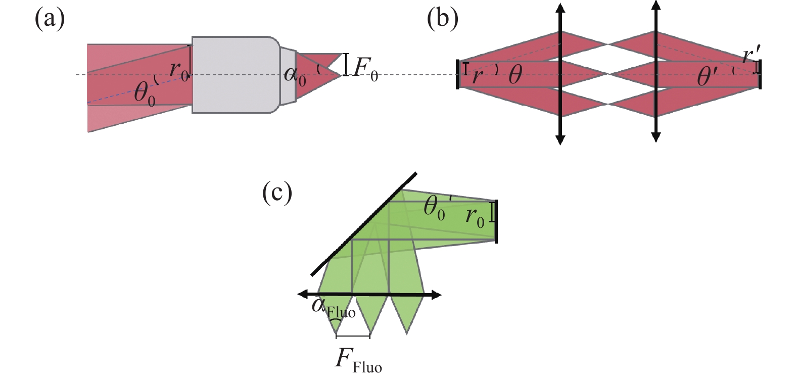
    Fig. 1. Definition of optical invariants. (a) Optical invariants of the imaging objectives; (b) Optical invariants of the scanning relay; (c) Optical invariants of the fluorescence collection system
    Design of a large-FOV two-photon microscope system using optical invariant analysis. (a) Optical layout; (b) Cerebral vasculature imaged over the mouse cortex with the large-FOV two-photon microscopy
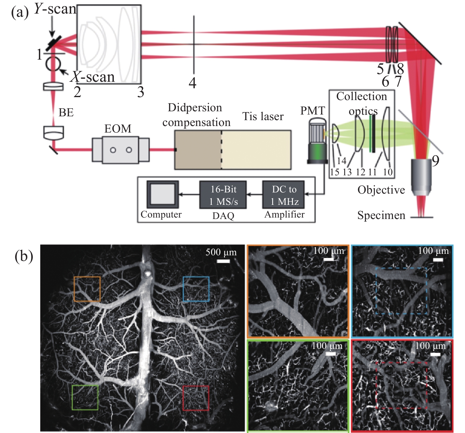
    Fig. 2. Design of a large-FOV two-photon microscope system using optical invariant analysis. (a) Optical layout; (b) Cerebral vasculature imaged over the mouse cortex with the large-FOV two-photon microscopy
    Large field-of-view two-photon microscopy by optimizing the off-axis aberrations using lens series. (a) Optical layout; (b) Experimental measurements of the PSF (point-spread-function) as a function of FOV, measured by 0.5 µm fluorescence beads; (c) Large-FOV two-photon image (λ= 800 nm, max intensity project) of vasomotion in cortical arterioles across both hemispheres of an awake, head-fixed mouse through dual transcranial windows
    Fig. 3. Large field-of-view two-photon microscopy by optimizing the off-axis aberrations using lens series. (a) Optical layout; (b) Experimental measurements of the PSF (point-spread-function) as a function of FOV, measured by 0.5 µm fluorescence beads; (c) Large-FOV two-photon image (λ= 800 nm, max intensity project) of vasomotion in cortical arterioles across both hemispheres of an awake, head-fixed mouse through dual transcranial windows
    Large-FOV two-photon random access microscopy. (a) Optical layout; (b) PSF at middle position and edge position of the FOV, measured by 0.5 µm microbeads; (c) Large-FOV two-photon image of fluorescent proteins in anesthetized thy-1 mice (max intensity project) and higher magnification image (dashed box in the large-FOV image)
    Fig. 4. Large-FOV two-photon random access microscopy. (a) Optical layout; (b) PSF at middle position and edge position of the FOV, measured by 0.5 µm microbeads; (c) Large-FOV two-photon image of fluorescent proteins in anesthetized thy-1 mice (max intensity project) and higher magnification image (dashed box in the large-FOV image)
    Large field-of-view and multi-region two-photon microscopy. (a) Optical layout; (b) PSF at middle position and edge position of the FOV, measured by 0.2 µm microbeads; (c) Large-FOV imaging was used to examine neuronal activity of a transgenic mouse expressing the genetically encoded fluorescent calcium indicator GCaMP6 s; (d) Segmenting the image sequence can yield 5,361 active neurons; (e) Simultaneous two region imaging to monitor neuronal activity between two cortical visual areas
    Fig. 5. Large field-of-view and multi-region two-photon microscopy. (a) Optical layout; (b) PSF at middle position and edge position of the FOV, measured by 0.2 µm microbeads; (c) Large-FOV imaging was used to examine neuronal activity of a transgenic mouse expressing the genetically encoded fluorescent calcium indicator GCaMP6 s; (d) Segmenting the image sequence can yield 5,361 active neurons; (e) Simultaneous two region imaging to monitor neuronal activity between two cortical visual areas
    Extending the field of view of two-photon microscopy using adaptive optics. (a) Schematic diagram; (b) Imaging optical path diagram; (c) Large-FOV two-photon image of the brain slice of thy-1 mice (max intensity project); (d) Comparison before and after adaptive optics correction in the yellow solid area of Fig. (c)
    Fig. 6. Extending the field of view of two-photon microscopy using adaptive optics. (a) Schematic diagram; (b) Imaging optical path diagram; (c) Large-FOV two-photon image of the brain slice of thy-1 mice (max intensity project); (d) Comparison before and after adaptive optics correction in the yellow solid area of Fig. (c)
    Jing Yao, Zhipeng Yu, Yufeng Gao, Shiwei Ye, Wei Zheng, Puxiang Lai. Advances of large field-of-view two-photon microscopy system (invited)[J]. Infrared and Laser Engineering, 2022, 51(11): 20220550
    Download Citation