• Acta Optica Sinica
  • Vol. 42, Issue 11, 1134011 (2022)
Jun Shi1, Yuxin Zhao1, Miao Li2、*, Feng Wang3, Guohong Yang3, and Minxi Wei3
Author Affiliations
  • 1Key Laboratory of Optoelectronic Technology and Systems, Ministry of Education, Chongqing University, Chongqing 400044, China
  • 2College of Optoelectronic Engineering, Chongqing University of Posts and Telecommunications, Chongqing 400065, China
  • 3Laser Fusion Research Center, China Academy of Engineering Physics, Mianyang 621900, Sichuan, China
  • show less
    DOI: 10.3788/AOS202242.1134011 Cite this Article Set citation alerts
    Jun Shi, Yuxin Zhao, Miao Li, Feng Wang, Guohong Yang, Minxi Wei. Hard X-Ray Transmission Imaging with Spherically Bent Crystal[J]. Acta Optica Sinica, 2022, 42(11): 1134011 Copy Citation Text show less
    Schematic diagrams of X-ray transmission and X-ray Bragg reflection. (a) Schematic diagram of X-ray transmission; (b) schematic diagram of X-ray Bragg reflection
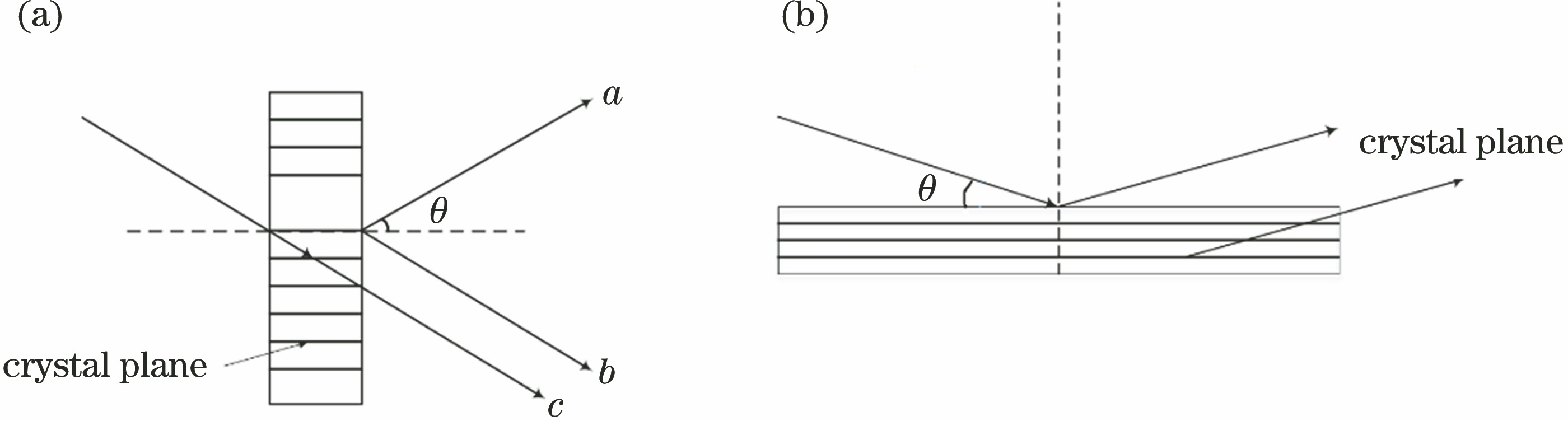
    Fig. 1. Schematic diagrams of X-ray transmission and X-ray Bragg reflection. (a) Schematic diagram of X-ray transmission; (b) schematic diagram of X-ray Bragg reflection
    Imaging optical path of spherical crystal. (a) Reflection imaging; (b) transmission imaging
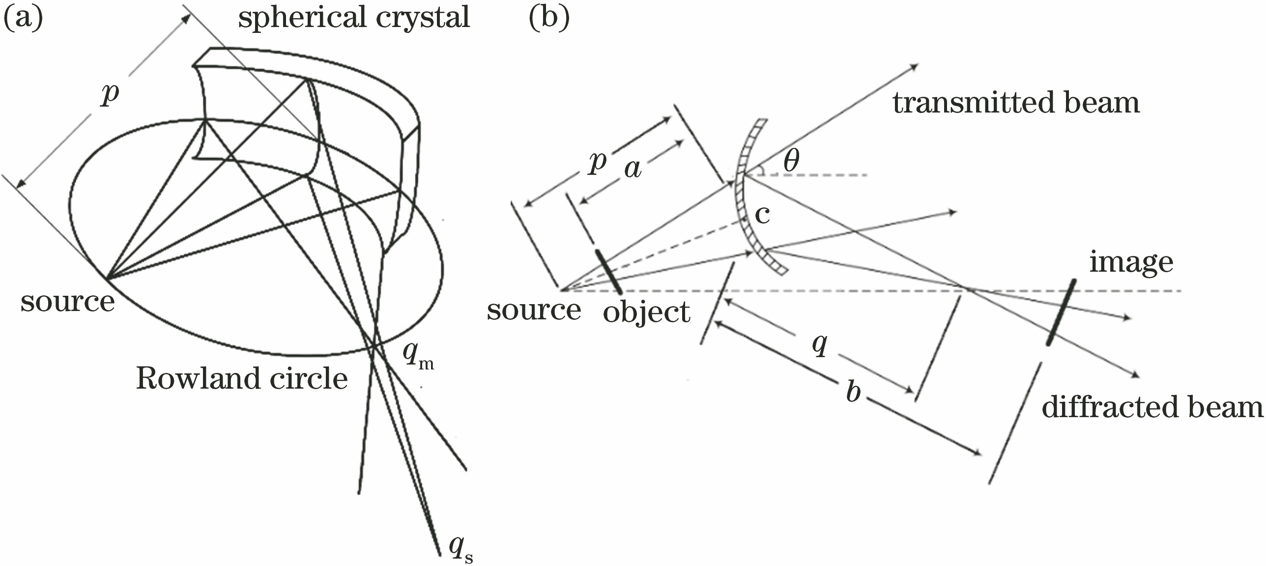
    Fig. 2. Imaging optical path of spherical crystal. (a) Reflection imaging; (b) transmission imaging
    Grid image results of crystal transmission simulation imaging under different b. (a) b=60.0 mm (Mm=0.74, Ms=1.87); (b) b=70.0 mm (Mm=0.86, Ms=1.93); (c) b=81.1 mm (Mm=1.00, Ms=2.00); (d) b=100.0 mm (Mm=1.23, Ms=2.12); (e) b=160.0 mm (Mm=1.97, Ms=2.50); (f) b=243.3 mm (Mm=3.00, Ms=3.00)
    Fig. 3. Grid image results of crystal transmission simulation imaging under different b. (a) b=60.0 mm (Mm=0.74, Ms=1.87); (b) b=70.0 mm (Mm=0.86, Ms=1.93); (c) b=81.1 mm (Mm=1.00, Ms=2.00); (d) b=100.0 mm (Mm=1.23, Ms=2.12); (e) b=160.0 mm (Mm=1.97, Ms=2.50); (f) b=243.3 mm (Mm=3.00, Ms=3.00)
    Picture of processed spherical transmission crystal
    Fig. 4. Picture of processed spherical transmission crystal
    Experimental diagram of Cu Kα backlight imaging with spherical transmission crystal
    Fig. 5. Experimental diagram of Cu Kα backlight imaging with spherical transmission crystal
    Results of spherical crystal transmission imaging under different b. (a) b=60 mm; (b) b=70 mm; (c) b=80 mm; (d) b=100 mm; (e) b=130 mm; (f) b=160 mm
    Fig. 6. Results of spherical crystal transmission imaging under different b. (a) b=60 mm; (b) b=70 mm; (c) b=80 mm; (d) b=100 mm; (e) b=130 mm; (f) b=160 mm
    CrystalMiller index2d /nmnEnergy /keVλ /nmθ /(°)
    Mica(002)1.984238--1000.0124--0.15501.074--13.553
    α-quartz(2020)0.426418--1000.0124--0.15501.666--21.316
    α-quartz(1011)0.668718--1000.0124--0.15501.063--13.403
    α-quartz(1010)0.851218--1000.0124--0.15500.835--10.492
    Ge(111)0.653218--1000.0124--0.15501.088--13.727
    Si(111)0.627118--1000.0124--0.15501.133--14.310
    Table 1. Bragg angle range in Laue transmission crystal structure
    Jun Shi, Yuxin Zhao, Miao Li, Feng Wang, Guohong Yang, Minxi Wei. Hard X-Ray Transmission Imaging with Spherically Bent Crystal[J]. Acta Optica Sinica, 2022, 42(11): 1134011
    Download Citation