
Cheng CHEN, Jingxin DING, Hui WANG, Deping WANG. Nd-doped Mesoporous Borosilicate Bioactive Glass-ceramic Bone Cement [J]. Journal of Inorganic Materials, 2022, 37(11): 1245
- Journal of Inorganic Materials
- Vol. 37, Issue 11, 1245 (2022)
Abstract
Keywords
As a malignant bone tumor, osteosarcoma is often found in adolescents, posing a serious threat to their health[1]. Most osteosarcoma can be treated by surgical resection, but it is difficult to prevent the recurrence and metastasis of residual tumor cells[2]. At the same time, the resection of osteosarcoma leads to serious bone defects, which seriously damage the health of patients[3]. Therefore, it is significant for the comprehensive treatment of osteosarcoma to develop biomaterials with both anti-tumor function and bone tissue repair function.
In recent years, due to the excellent biological activity and degradation properties, bioactive glass bone cement has been widely studied in bone defect repair[4]. Bioactive glass bone cement is prepared by mixing bioactive glass powder and the liquid phase, and then injected into the bone defect site by a minimally invasive method. It provides mechanical support for the early self-healing period after solidification. Meanwhile, the functional ions released from bioactive glass can effectively promote the regeneration of bone tissue[5-6]. In our previous research, borosilicate bioactive glass with controllable degradation properties was prepared by replacing part of SiO2 of silicate bioactive glass by B2O3, and the divalent cations released by the degradation of borosilicate bioactive glass were chelated with liquid alginate, which enhanced the handling performance and biological activity of bone cement[7⇓-9]. In addition, we have reported that mesoporous borosilicate bioactive glass microspheres were loaded with drug molecules and used as smart carriers for local drug slow-release[10].
In addition to repairing bone defects, killing residual tumor cells is also an indispensable task in the postoperative treatment of osteosarcoma. Photothermal therapy (PTT) and chemotherapy (CHT) are common adjuvant treatments after tumor surgery, and the combination of the two has been proven to have certain advantages in improving tumor treatment efficiency and reducing side effects[11⇓-13]. This is because the heat generated by PTT can both kill tumor cells and accelerate the entry of drug molecules into tumor cells[11]. In addition, tumor cells are more sensitive to chemical drugs when heated, which helps to improve the treatment efficiency of anti-cancer drugs[14]. Yang et al.[15] synthesized a redox-responsive drug delivery system, which effectively inhibited the growth of lung cancer cells through photothermal- chemotherapy function. Zhao et al.[16] developed a hollow polymer-silica nanohybrid as a nanocarrier to simultaneously deliver the photothermal agent and anti- cancer drug to tumors for chemo-photothermal synergistic therapy, which achieved better therapeutic effects in the treatment of high-grade malignant tumor models. However, there are currently few reports on the research of PTT&CHT combination therapy for osteosarcoma. So it deserves to try to use the combination therapy to improve the treatment effect of osteosarcoma.
Among rare earth ions, Nd3+ with excellent photothermal properties, can be effectively excited by the 808 nm laser located in the first biological window and has a larger absorption cross section[17-18]. With increasing doping concentration, Nd3+ has a concentration quenching effect, which makes heavily neodymium-doped nanoparticles contain a highly light-to-heat conversion efficiency[19]. At the same time, Nd3+ doped biomaterials show good biocompatibility[20-21].
Accordingly, in this study, we first prepared neodymium-doped mesoporous borosilicate bioactive glass- ceramic (MBGC-xNd) microspheres with good photothermal properties through the Sol-Gel method combined with solid-state reaction[22], and used it for loading an anticancer drug doxorubicin (DOX). Subsequently, as solid phase, MBGC-xNd microspheres were mixed with sodium alginate (SA) solution to obtain a multifunctional bone cement (MBGC-xNd/SA) for regeneration of bone tissue and synergistic therapy of osteosarcoma (Scheme 1). The microscopic morphology, phase composition and mesoporous structure of MBGC-xNd microspheres were characterized. The photothermal properties and DOX release behavior of microspheres and bone cement were characterized, and setting properties of bone cement were tested. The biological activity of promoting proliferation and differentiation of rabbit bone mesenchymal stem cells (rBMSCs) of the prepared bone cement were preliminarily evaluated in vitro. In addition, the killing effect of PTT&CHT combination therapy on osteosarcoma cells (MG-63) was also evaluated.
![]()
Figure 1.Characterization of MBGC-
1 Materials and Methods
1.1 Materials
Tri-n-butyl borate (TBB), neodymium nitrate hexahydrate (Nd(NO3)3·6H2O), calcium nitrate tetrahydrate (Ca(NO3)2·4H2O), tetraethyl orthosilicate (TEOS), ammonia solution (25 % NH3 in water, in mass), ethyl acetate, ethanol absolute, triethylphosphate (TEP), doxorubicin hydrochloride, cetyl trimethyl ammonium bromide (CTAB), sodium alginate (SA), gluconolactone, and Na2HPO4 were purchased from Sinopharm Chemical Reagent Co., Ltd (Shanghai, China). The BCA protein concentration determination kit, fetal bovine serum (FBS), dexamethasone, alkaline phosphate assay kit, beta-glycerophosphate disodium salt, and Vitamin C were purchased from Beyotime Biotechnology (Shanghai, China). Low-glucose dulbecco's modified eagle medium (DMEM), high-glucose DMEM, 1% penicillin/streptomycin, and CCK-8 reagent were purchased from Titan Scientific Co., Ltd (Shanghai, China). MG-63 cells and rBMSCs were obtained from Shanghai Sixth People’s Hospital.
1.2 Preparation of MBGC-xNd microspheres
The mesoporous SiO2-B2O3-P2O5 nanospheres (MBN) were prepared through the Sol-Gel method[23], and their chemical composition (in mol) was 78% SiO2+16% B2O3+6% P2O5. Firstly, 0.7 g CTAB and 20 mL ethyl acetate were added into 40 mL deionized water under stired at 30 ℃. 30 min later, 0.7 mL ammonia solution were added. Later, 2.43 mL TEOS, 1.67 mL TBB and 0.288 mL TEP were added every 30 min. After 4 h, the reaction solution was centrifuged to collect the precipitate. After washed three times by absolute ethanol and deionized water, the precipitate was vacuum dried at 37 ℃ to obtain MBN.
MBGC-xNd microspheres were prepared via solid- state reaction[22]. The composition of MBGC-xNd microspheres is shown in Table 1, where Nd3+ replaced Ca2+ in an equimolar ratio (x=0, 0.01, 0.03, 0.05). The specific process was as follows. A certain amount of calcium nitrate tetrahydrate and neodymium nitrate hexahydrate was dissolved in 40 mL absolute ethanol. Then MBN was added, and the suspension was stirred until absolute ethanol was completely volatilized. The collected powder was annealed at 600 ℃ for 5 h to prepare MBGC-xNd microspheres.
| Sample | SiO2 | B2O3 | P2O5 | CaO | NdO3/2 |
|---|---|---|---|---|---|
| MBGC-0Nd | 50 | 10 | 4 | 36 | 0 |
| MBGC-1Nd | 50 | 10 | 4 | 35 | 1 |
| MBGC-3Nd | 50 | 10 | 4 | 33 | 3 |
| MBGC-5Nd | 50 | 10 | 4 | 31 | 5 |
Table 1.
Composition in molar percent of MBGC-xNd microspheres
1.3 Preparation of MBGC-xNd/SA
2 g SA, 1 g gluconolactone and 3 g Na2HPO4 were dissolved in 100 mL deionized water to form a solution, which, as the liquid phase, was mixed and stirred with MBGC-xNd microspheres at a liquid-solid ratio of 1.3 mL/mg. After stirred for 60 s, the bone cement slurry was poured into a ϕ6 mm×12 mm polytetrafluoroethylene cylindrical mold for molding, then stored at 37 ℃ for 24 h.
1.4 Characterization of MBN, MBGC-xNd microspheres and MBGC-3Nd/SA
The microscopic morphology of MBN and MBGC-xNd microspheres was observed by transmission electron microscope (TEM; H-800; Japan). The phase composition of MBN and MBGC-xNd microspheres was tested by X-ray polycrystalline diffractometer (XRD; Rigaku SmartLab 9; Japan). The mesoporous parameters of MBN and MBGC-xNd microspheres were tested by specific surface area analyzer (BET; Autosorb-Nova 2200 e; America). After dried and sprayed with gold, the morphology of MBGC-3Nd/SA was observed via scanning electron microscope (SEM; S-4700; Japan).
1.5 Characterization of degradation performance
The degradation performance of MBGC-xNd microspheres was characterized by measuring the change of Ca and Si concentrations in simulated body fluid (SBF). MBGC-xNd microspheres were soaked in SBF at pH 7.4 (1 mg/mL), then stored at 37 ℃. After 1, 3, 7, and 10 d, Ca and Si concentrations were tested through inductively coupled plasma emission spectrometer (ICP; PE 8300; America).
1.6 Drug loading and release
1.6.1 Drug loading of MBGC-xNd microspheres
Firstly, 0.25 mg/mL DOX-PBS solution was configured by dissolving doxorubicin hydrochloride in phosphate buffer solution (PBS). Then 50 mg MBGC-xNd microspheres were added in 6 mL DOX solution. After shaking for 1 d, the solution was centrifuged, the supematant was collected, and precipitated MBGC-xNd@DOX microspheres were obtained and dried at 37 ℃. Next, DOX standard solutions (0.006-0.06 mg/mL) were configured, and their DOX concentration was tested by ultraviolet-visible spectroscope (UV-Vis; U-4100; Japan) at an absorption wavelength of 480 nm[23]. The obtained standard curve equation between the measured standard concentration of DOX and the absorbance value was as follows.
(R2=0.9992), where A is the absorbance value, C is the DOX concentration.
Absorbance value of the above-mentioned centrifuged supernatant was measured, then substituted into the Eq.(1) to obtain the concentration of residual DOX in the solution. Finally, the DOX encapsulation efficiency (E) was calculated by Eq. (2)[23]:
Where Ct is the initial DOX concentration, and Cr is the residual DOX concentration.
1.6.2 Drug release from MBGC-xNd@DOX microspheres
The dried MBGC-xNd@DOX microspheres were soaked in 7 mL PBS at pH 4.7, and shaken at 120 r/min in a 37 ℃ shaker. After shaking for 1, 3, 6, 9, 12, 24, 48, 72, 168, and 240 h, 3.5 mL solution was aspirated to measure the DOX concentration and new PBS was supplemented. The cumulative concentration of DOX released from MBGC- xNd@DOX microspheres was calculated by Eq. (3)[24]:
Where Ct is the DOX concentration of the solution at time t (t = 1, 3, 6, 9, 12, 24, 48, 72, 168, and 240 h). Five parallel experiments were tested and the average DOX cumulative concentration was calculated.
1.6.3 Drug release from MBGC-xNd/SA drug- loaded bone cement
MBGC-xNd/SA drug-loaded bone cement (MBGC- xNd/SA@DOX) was obtained by mixing MBGC-xNd@DOX microspheres and sodium alginate solution. The prepared MBGC-xNd/SA@DOX (6 mm×6 mm×3 mm) were immersed in 7 mL PBS at pH 4.7 for the drug release experiment. For characterizing the influence of temperature on the DOX release behavior, the temperature of the shaker was adjusted to 35, 45 and 55 ℃ to carry out the drug release experiment of MBGC-3Nd/ SA@DOX.
1.7 Characterization of photothermal properties
For studying the influence of Nd3+ doping amount on photothermal properties of MBGC-xNd microspheres, 3.2 W/cm2 808 nm laser was used to irradiate MBGC- xNd microspheres with different Nd3+ doping amounts for 5 min with an on and off cycle for five times. The MBGC-0Nd microsphere was used as a control sample. An infrared thermal camera system (Fluke, Everett, WA, America) was used to record temperature changes in time. In addition, different power densities of 808 nm lasers (0.56, 0.99, 1.5, and 3.2 W/cm2) were used for irradiating MBGC-3Nd microspheres to characterize the influence of power densities of laser on the photothermal properties of microspheres.
Furthermore, bone cement samples were soaked in 10 mL SBF and irradiated under 2.4 W/cm2 laser to characterize the photothermal properties of MBGC-0Nd/SA and MBGC-3Nd/SA after five switching cycles.
1.8 Setting properties of MBGC-xNd/SA
1.8.1 Setting time measurement
The setting time was recorded according to ISO 9917-1. The bone cement slurry was prepared by above method and poured into a ϕ6 mm×12 mm polytetrafluoroethylene cylindrical mold. The bone cement slurry was considered to be set when the needle (1.13 mm in diameter, 400 g in weight) of the Vicat instrument fell freely on the surface of the bone cement and failed to cause obvious indentation (indentation depth≤0.5 mm). The setting time was defined as a period from the preparation of the slurry to the set of the slurry. The average setting time was calculated by testing five parallel experiments.
1.8.2 Injectability measurement
Bone cement slurry was squeezed out from a syringe (1.7 mm in diameter) at a loading rate of 15 mm/min until the indenter pressure reached 100 N. Injectability (Inj.) was calculated as follows:
Where W0 is the weight of the empty syringe, W1 is the weight of the syringe containing bone cement slurry, and W2 is the weight of the syringe containing the residual bone cement slurry. The average injectability was calculated by testing five parallel experiments.
1.8.3 Compressive strength measurement
The bone cement samples cured for 24 h in a box with constant temperature and humidity were taken out, and the surface moisture was wiped with filter paper. After the upper and lower surfaces of the samples were polished with sandpaper into parallel surfaces, the samples were placed in an electronic universal testing machine (CTM250; China), and loaded at 0.5 mm/min loading rate to measure compressive strength. The average compressive strength was calculated by testing five parallel experiments.
1.8.4 Anti-washout properties measurement
After curing for 24 h and drying, bone cement samples were soaked in PBS at pH 7.4 (0.2 g/mL), then placed in a 37 ℃ gas bath thermostat and oscillated at the rate of 180 r/min. After oscillating for 6, 12, 18 and 24 h, the samples were taken out and dried. The anti-washout properties were calculated by Eq. (5):
Where R. is the remaining weight ratio, W0 is the initial dry weight of the bone cement, Wt is the remaining dry weight of the bone cement at time t (t=6, 12, 18, and 24 h). The average value was calculated by testing five parallel experiments.
1.9 Cell viability test of rBMSCs
1.9.1 Test of cell proliferation
BMSCs were cultured in low-glucose DMEM containing 10% FBS and 1% penicillin/streptomycin in cell incubator (ESCO, Singapore) in condition of 37 ℃ and 5% CO2. The bone cement samples were prepared to a size of ϕ6 mm×3 mm for in vitro cell experiments. The samples were autoclaved and immersed in low-glucose DMEM (100 mg/mL). After 24 h, the extract of the bone cement samples was collected through filter membrane and diluted 40 times with low-glucose DMEM. The rBMSCs were seeded in 96-well plates (500 cells/well) and cultured in low-glucose DMEM for 1 d. Next, the diluted DMEM containing extract was added to replace the previous medium and replaced every two days. After culturing for 1, 3 and 5 d, the OD value was tested by CCK-8 reagent. The average OD value was calculated by testing five parallel experiments.
1.9.2 Test of alkaline phosphatase (ALP) activity
The osteogenic differentiation ability was evaluated by measuring ALP activity of rBMSCs cultured in the bone cement extract[25]. The bone cement extract was diluted 40 times with low-glucose DMEM, then added with a final concentration of 10 mmol/L β-glycerophosphate disodium salt, 0.1 μmol/L dexamethasone, and 50 μmol/L Vitamin C to configure the osteogenic differentiation medium. The rBMSCs were seeded in 48-well plates (2×10⁴ cells/well) and cultured in low-glucose DMEM for 1 d. Then the osteogenic differentiation medium was added to replace the previous medium and replaced every 2 d. The ALP activity was evaluated on the 7th and 14th days by alkaline phosphate assay kit. After staining for 30 min, the optical density (OD) value was tested at 405 nm. At the same time, the cell protein content in each well was tested through the BCA protein concentration determination kit. The OD value divided by the total cell protein content to calculate the relative ALP activity. The average value was calculated by testing five parallel experiments.
1.10 Anti-tumor experiment of PTT&CHT combination therapy
MG-63 osteosarcoma cells were cultured in high- glucose DMEM containing 10% FBS and 1% penicillin/ streptomycin in cell incubator in condition of 37 ℃ and 5% CO2. Then MG-63 cells were seeded in 48-well plates (2×10⁴ cells/well). After 1 d of incubation, the autoclaved MBGC-0Nd/SA, MBGC-3Nd/SA, and MBGC- 3Nd/SA@DOX were placed in 48-well plates, and each group of bone cement samples was divided into the laser irradiation group and the non-laser irradiation group. MBGC-0Nd/SA without laser irradiation was used as a blank control group. MBGC-3Nd/SA with laser irradiation, MBGC-3Nd/SA@DOX without laser irradiation and MBGC-3Nd/SA@DOX with laser irradiation were labeled as PTT group, CHT group and CHT&PTT combination therapy group. Then the laser irradiation group was irradiated under 2.4 W/cm2 808 nm laser for 5 min. After co-culturing for 12 h, the previous medium and bone cement were removed, and 50 μL CCK-8 reagent and 450 μL high-glucose DMEM were added. Two hours later, the OD value was tested. Finally, the OD value of the blank control group was regarded as 100%, and the proportion of the OD value of the other groups accounting for the OD value of the blank control group was calculated to obtain the tumor cell survival rate. The average value was calculated by testing five parallel experiments.
1.11 Statistical analysis
The experimental results were represented as mean± standard deviation, and statistical analysis was performed via one-way ANOVA. When p<0.05 (*), the data were considered significantly different.
2 Results and discussion
2.1 Characterization of MBGC-xNd microspheres
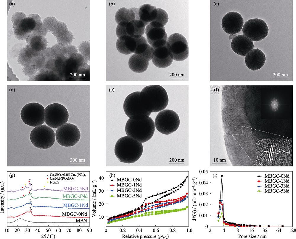
As shown in Fig. 1(a-e), all microspheres were in regular spherical shape with diameters about 250 nm, and displayed obvious slit-type mesoporous structure, which was attributed to the removal of the templating agent CTAB. Compared with MBN, the internal structure of MBGC-xNd microspheres was more compact. It showed that the morphology and size of MBN were not significantly affected by the introduction of Ca2+ and Nd3+ through solid-state reaction, but the mesoporous structure of microspheres was affected to some extent (Fig. 1(f)).
![]()
Figure 1.Characterization of MBGC-
Fig. 1(g) showed the phase composition of MBN and MBGC-xNd microspheres after heat treatment at 600 ℃. It obviously exhibits broad peaks existed in all samples, which were attributed to the glass phase structure of MBN, while the solid-state reaction of introducing Ca2+ and Nd3+ produced a clear crystalline phase composition. All MBGC-xNd microsphere data showed diffraction peaks at 2θ=32.4° and 33.2°, which were in line with the diffraction characteristic peaks of Ca2SiO4·0.05Ca3(PO4)2 (PDF#49-1674). However, the data of MBGC-3Nd and MBGC-5Nd microspheres showed diffraction peaks at 2θ=25.8° and 31.8°, which were in line with the diffraction characteristic peaks of Ca8Nd2(PO4)6O2 (PDF#32- 0175). Especially, the diffraction characteristic peaks of Nd2O3 (PDF#65-3184) appeared in MBGC-5Nd microsphere data at 2θ=27.9° and 46.3°. In order to prevent Nd3+ with high ionic potential from destroying the balance of the microemulsion system, the solid-state reaction was used to incorporate Ca2+ and Nd3+ into MBN. Therefore, it can be considered that Ca2+ and Nd3+ were deposited on the surface and pores of MBN by adsorption. Ca2+ and Nd3+ gradually diffused into the glass network and competed for non-bridging oxygen during the sintering process, causing an increase in free energy of the system, promoting the formation of crystalline phases[26]. In addition, the results also showed that the final crystalline phase composition might be affected by the relative content of Ca2+ and Nd3+.
As can be seen from Fig. 1(f), the amorphous morphology existed in the MBGC-3Nd microspheres, and the diffraction fringes of the crystals were also distributed in the microspheres. The interplanar crystal spacing was around 0.334 nm, which was consistent with the lattice spacing of (002) plane of Ca8Nd2(PO4)6O2. The results showed that the crystal phase generated during the solid- state reaction existed in the glass phase in the form of dispersed crystallites. Type IV isotherm and H3 hysteresis loop were seen in all N2 adsorption-desorption isotherms of MBGC-xNd microspheres, revealing the slit- type mesoporous structure of microspheres (Fig. 1(h)). However, with the increase of Nd3+ content, the hysteresis loop of MBGC-xNd microspheres got smaller, demonstrating the reduction of specific surface area. The pore size distribution range of MBGC-xNd microspheres was narrow, mainly in the range of 3-8 nm (Fig. 1(i)).
As shown in Table 2, compared with MBN, the specific surface area, pore size, and pore volume of MBGC-xNd microspheres reduced. This is because Ca2+ and Nd3+ were deposited on the surface and pores of MBN by adsorption, and gradually diffused into the glass network during the sintering process. Therefore, there might be some formed microcrystalline phase stacking in the pores, resulting in a decrease in these mesoporous parameters[22]. In addition, the pore size of MBGC-xNd microspheres decreased with the increase of Nd3+ content. When Nd3+ doping concentration was low, it replaced Ca2+ as network modifier[27-28]. The ionic radii of Nd3+ and Ca2+ were similar, but the charge of Nd3+ was higher. Therefore, Nd3+ with higher ionic potential might enable the glass network tighter, resulting in the shrinkage of the mesoporous structure.
| Sample | Specific surface | Pore size /nm | Pore volume |
|---|---|---|---|
| MBN | 329.261 | 12.506 | 0.4498 |
| MBGC-0Nd | 42.092 | 6.024 | 0.0634 |
| MBGC-1Nd | 36.065 | 4.776 | 0.0431 |
| MBGC-3Nd | 34.424 | 4.496 | 0.0387 |
| MBGC-5Nd | 28.818 | 3.710 | 0.0268 |
Table 2.
Pore structure of MBN and MBGC-xNd microspheres
Bioactive glass in the body fluid environment can release functional ions (such as Ca2+ and SiO44-), which play a significant role in promoting bone regeneration[4,7]. MBGC-xNd microspheres showed obvious degradation in SBF by releasing Ca and Si (Fig. 2). At the early stage, the Si and Ca concentrations increased rapidly, then Si concentration remained relatively stable and Ca concentration even decreased. It is because a large amount of free SiO44- and Ca2+ on the surface of the microspheres were released into the solution at the initial stage. As the Si concentration reached saturation, SiO44- began to accumulate on the surface to form a silicon-rich layer[7]. Ca2+ reacted with PO43- in the solution to form hydroxyapatite deposited on the surface, consuming the Ca2+ in the solution[29]. Besides, the ionic degradation rate of MBGC-xNd decreased with the rise of Nd3+ content, which was attributed to the progressively strengthened glass network. On the one hand, with the reduction of specific surface area, the contact area between microspheres and SBF decreased, and on the other hand, it was difficult in releasing ions.
![]()
Figure 2.Changes of (A) Ca and (B) Si concentrations with MBGC-
Adequate drug loading content and sustained drug release behavior are essential for effective chemotherapy. The results showed the encapsulation efficiency of MBGC-0Nd, MBGC-1Nd, MBGC-3Nd, and MBGC-5Nd microspheres was 89.2%, 84.6%, 82.3%, and 73.8%, respectively. As can be observed in Fig. 3(A), all MBGC- xNd@DOX microspheres showed the sustained drug release behavior. As the Nd3+ content increasing, the total amount of DOX released during the whole period decreased from about 45% (MBGC-0Nd) to about 35% (MBGC-5Nd). It is due to the influence of Nd3+ content on the mesoporous structure of microspheres, and the decrease of these mesoporous parameters resulted in the reduction of DOX encapsulation efficiency and the difficulty in drug release. In addition, for such mesoporous microspheres, DOX is mainly loaded by mesoporous through physical adsorption[10]. However, the degradation of microspheres is caused by ion exchange between the glass network and surrounding solution. Therefore, DOX based on physical adsorption exerted less effect on the degradation process based on chemical action. However, the accumulative amount of DOX released from MBGC-3Nd during the whole period could increase with the temperature rising and attain more than 55% Fig. 3(B), which obviously indicated that temperature might be used as an effective way to control the release.
![]()
Figure 3.Cumulative release curves of DOX from (A) MBGC-
The photothermal properties of MBGC-xNd microspheres were characterized under 808 nm near-infrared laser irradiation. The microspheres doped with Nd3+ showed a rapid rise of temperature under 3.2 W/cm2 808 nm laser irradiation, and maintained a stable heating effect during the five switching cycles of the laser (Fig. 4(A-C)),while MBGC-0Nd microspheres did not show the photothermal effect (Fig. 4(A)). Meanwhile, the final temperature of MBGC-xNd microspheres rose with the increase of Nd3+ content. Nd3+ ([Xe]4f3) has particular electronic configuration with a half-filled 4f orbit. As Fig.4(D) showed, electrons are excited to an excited state (4F5/2) under the laser irradiation of 808-nm, then rapidly return to the metastable ground state 4F3/2 by the nonradiative process. Later, Nd3+ undergoes a radiative decay to lower energy states (such as 4F13/2, 4F11/2 and 4F9/2), and the phonon-assisted decay occurs in the ground state, which results in photothermal effect[30⇓-32]. In view of lower Nd3+ content and better photothermal properties, MBGC-3Nd microspheres were chosen for further experiments (Fig. 4(C)). With the increase of power density, a growing tendency was represented in the final temperature of MBGC-3Nd microspheres under the irradiation of 808 nm laser (Fig. 4(B)).
![]()
Figure 4.Photothermal properties of MBGC-
2.2 Characterization of MBGC-3Nd/SA bone cement
The prepared MBGC-3Nd/SA bone cement showed excellent injectability (Fig. 5(A)). The MBGC-3Nd microspheres were coated with sodium alginate to prepared MBGC-3Nd/SA and formed a whole through cross-linking. However, the bone cement was not completely dense, and there were some gaps between the clusters formed by the cross-linking of the microspheres and alginate, which might facilitate the drug release from the bone cement (Fig. 5(B)). As shown in Fig. 5(C), when MBGC-3Nd microspheres were in contact with the liquid phase, the surface glass network was destroyed, and Ca2+ were released from the surface, which then cross-linked with alginate on the glass surface to form the Ca-Alg gel[7⇓-9]. With the continuous dissolution of Ca2+, the generated Ca-Alg gel gradually wrapped the microspheres like a packaging bag to form a whole, and gradually realized the coagulation and solidification of the bone cement. Moreover, previous studies have shown that borates can be complexed with diols in the sugar ring of alginate, leading to formation of mono- and di-complexes[33-34]. This indicated that the B component might play a prominent role in the cross-linking process of MBGC-3Nd microspheres and alginate.
![]()
Figure 5.Characterization of MBGC-3Nd/SA bone cement(A) Photo of extruded bone cement; (B) SEM image of MBGC-3Nd/SA; (C) Schematic illustration of the setting process of MBGC-3Nd/SAThe color figures can be obtained from online edition
The influence of Nd3+ content on the setting properties of MBGC-xNd/SA is shown in Fig. 6. With the increase of Nd3+ content, the setting time of bone cement increased from about 27 min to about 34 min (Fig. 6(A)). The reason lied in the crosslinking speed between MBGC microspheres and alginate depends on the dissolution speed of Ca2+, which gradually decreases with the increase of Nd3+ content, resulting in prolonged setting time of bone cement. In addition, the cumulative dissolution concentration of Ca2+ of microspheres also decreased with the increase of Nd3+ content, resulting in insufficient cross-linking with alginate and a decrease in the compressive strength of bone cement (Fig. 6(C)). As shown in Fig. 6(B), more than 90% of the bone cement slurry was extruded from the syringe within 2 min, and there was no significant difference in the injection rate of all bone cement samples, indicating that the incorporation of Nd3+ exerted no significant effect on the injectability of bone cement. It can be seen from Fig. 6(D) that with the rise of Nd3+ content, the remaining mass of samples reduced, indicating the anti-washout property gradually weakened. However, the remaining mass of all samples was more than 90%, demonstrating the good anti-washout property. In summary, MBGC-xNd/SA performed good operability and suitable for clinical minimally invasive surgery.
![]()
Figure 6.Setting properties of MBGC-
As shown in Fig. 4(C), the temperature of MBGC- 3Nd/SA immersed in SBF increased rapidly under 2.4 W/cm2 808 nm laser irradiation and reached about 53 ℃, then maintained a stable heating effect within five switching cycles of the laser. According to previous studies, the suitable temperature for photothermal therapy is about 53 ℃, which not only effectively inhibits tumor growth, but also causes less damage to tissues[20,35]. Meanwhile, MBGC-0Nd/SA did not exhibit obvious photothermal properties under the same laser irradiation. The results show that the photothermal properties of MBGC-3Nd were not affected by the alginate composition. In addition, at different ambient temperatures, the sustained drug release behavior of MBGC-3Nd/SA@DOX was exhibited, and the cumulative release of DOX increased with the rise of ambient temperature, which may be the result of faster diffusion of DOX at higher temperature (Fig. 3(B)). This result demonstrates the heat generated by the photothermal therapy possessed the possibility of promoting the release of DOX. According to the previous studies, the IC50 data of DOX on MG-63 osteosarcoma cells was 3.87 μg/mL[36], while in the high DOX concentration of 50 μg/mL, the survival rate of rat mesenchymal stem cells after 48 h culturing was still higher than 50%[37]. This indicates that the cytotoxicity of DOX to osteosarcoma cells is much higher than to mesenchymal stem cells. Therefore, it is possible to avoid photothermal-promoted release of DOX to destroy the osteogenic function by adjusting the drug loading and the laser irradiation time.
Fig. 7 showed the effects of MBGC-0Nd/SA and MBGC-3Nd/SA extracts on the osteogenic properties of rBMSCs. As can be seen from Fig. 7(A), compared with the blank control, rBMSCs cultured in MBGC-0Nd/SA and MBGC-3Nd/SA bone cement extracts showed higher viability on the fifth day, reflecting their effect of promoting cell proliferation. Meanwhile, no significant difference was seen in the proliferation behavior of rBMSCs between MBGC-0Nd/SA and MBGC-3Nd/SA extracts, indicating that the cytocompatibility of bone cement was not affected by the incorporation of Nd3+. Furthermore, the alkaline phosphate (ALP) activity of rBMSCs was shown to be stimulated by extracts of MBGC-0Nd/SA and MBGC-3Nd/SA (Fig. 7(B)). These results suggest that the MBGC-xNd/SA bone cement possesses good cytocompatibility and bioactivity to promote the proliferation and expression of ALP activity of rBMSCs. Previous studies have also shown that borosilicate bioactive glass bone cement has good biocompatibility and can promote new bone regeneration in rabbit femoral condyle defects[38]. It is thus reasonable to believe that the MGBC-xNd bone cement might be used as a bioactive material to repair bone defects.
![]()
Figure 7.(A) Proliferation results and (B) alkaline phosphate activity of rBMSCs cultured in the bone cement extract*:
Previous studies have shown that benefiting from the release of active ions such as SiO44- and Ca2+, which can promote the formation of hydroxyapatite, borosilicate bioactive glass has good bioactivity and can promote bone tissue regeneration[39⇓⇓-42]. In addition, borate with low concentrations can stimulate cell growth and proliferation by activating the MAPK pathway[43]. Recently, Ma et al.[20] reported that Nd-doped bioglass has high bioactivity in stimulating angiogenesis, which improves the ability to promote tissue regeneration. It is thus reasonable to believe that MBGC-xNd/SA might be a bioactive material which could promote the regeneration of bone tissue. However, the mechanisms of Nd3+ in regulating cellular activity need to be investigated by further studies.
As shown in Fig. 8(A), the relative survival of co- cultured MG-63 cells decreased from (95.82±3.81)% to (25.15±2.35)% after MBGC-3Nd/SA was irradiated by 2.4 W/cm2 808 nm laser for 5 min, while no significant difference was seen in the relative survival of MG-63 cells co-cultured with MBGC-0Nd/SA before and after the laser irradiation. This result indicated that the photothermal therapy of MBGC-3Nd/SA had obvious killing effect on MG-63 cells. Furthermore, compared with PTT or CHT, the relative survival of MG-63 cells under combination therapy was significantly lower, only (7.04±3.32)%, indicating the synergistic effect of PTT&CHT combination therapy (Fig. 8(B)). Indeed, MG-63 cells are human osteosarcoma cells, and it possesses the property of both osteoblast like cells and tumor like cells. Furthermore, it also can differentiate into osteoblasts under the induction of growth factor. Due to the poorer heat dissipation capacity, tumor tissue has been shown to be more thermosensitive than normal tissue[44-45]. Therefore, during the anti-tumor treatment state, PTT can selectively kill the tumor cells with harmless effects on the normal cells. In addition, according to our previous studies, localized pH rises with the degradation of borosilicate bioactive glass[7]. Such a slightly alkaline environment can promote the proliferation of osteoblasts[46] but inhibit the proliferation of tumor cells.
![]()
Figure 8.Relative survival of MG-63 cells co-cultured with (A) bone cement under 808 nm laser irradiation at a power density of 2.4 W/cm2 for 5 min and (B) MBGC-3Nd/SA under PTT, CHT or combination therapy.*:
In recent years, due to the efficacy and selectivity, PTT has received much attention in bone tumor therapy. Ma et al.[47] fabricated a graphene oxide-β-tricalcium phosphate composite stent by 3D printing and surface modification strategy, which combined significantly improved osteogenic capacity with a high photothermal effect. Chen et al.[48] developed a novel functional tricalcium silicate bone cement with excellent photothermal properties for minimally invasive treatment of bone tumors. Liu et al.[49] prepared Mn-doped mesoporous bioactive glass as a phototherapy agent for bone regeneration and bone tumor treatment. However, PTT has a major shortcoming: the heat applied to the tumor is often uneven, leading to some cancer cells escape to survive and recurrence[16,50]. Therefore, it is significant to combine PTT with other therapy methods to improve the therapy efficiency of bone tumor. As we all know, CHT is a traditional therapy method for cancer treatment that can significantly inhibit the tumor growth rate. Previous studies have shown that the combination of PTT and CHT achieved a significant synergistic effect[51⇓-53]. In this study, the developed MBGC-3Nd/SA bone cement not only maintained the good bioactivity of borosilicate bioactive glass-ceramics, but also performed simultaneous photothermal therapy and chemotherapy on osteosarcoma cells by using MBGC-3Nd microspheres as photothermal agents and anti-cancer drug carriers. The combination therapy of PTT and CHT showed a significant synergistic effect on osteosarcoma cells, which improved the therapy efficiency. Hence, the combination of PTT and CHT is a potential therapy strategy with high efficacy for the synergistic therapy of osteosarcoma.
3 Conclusion
In this work, a Nd-doped mesoporous borosilicate bioactive glass-ceramic bone cement (MBGC-xNd/SA) for repair of bone defects and synergistic therapy of osteosarcoma is prepared by mixing Nd-doped mesoporous borosilicate bioactive glass-ceramic (MBGC-xNd) microspheres and sodium alginate (SA) solution. The results demonstrate that Nd3+ endowed MBGC-xNd microspheres with controllable photothermal properties, which can be adjusted by changing Nd3+ content and the power density of NIR laser. MBGC-xNd microspheres loaded with DOX all show sustained drug release behavior, but the mesoporous structure of MBGC-xNd microspheres shrinks with the incorporation of Nd3+ content, resulting in a slower degradation rate and a lower DOX encapsulation efficiency. The prepared MBGC-3Nd/SA bone cement exhibits good photothermal properties, and the heat generated by the photothermal therapy has the possibility of promoting the release of DOX. In addition, MBGC-3Nd/SA exhibits osteogenic bioactivity of promoting the proliferation and the expression of alkaline phosphatase activity of rBMSCs in vitro. More importantly, the synergistic effect of photothermal-chemical combination therapy of MBGC-3Nd/SA is demonstrated in tumor cell experiments. All above results consolidate that MBGC-3Nd/SA can be a promising material for postoperative treatment of osteosarcoma.
References
[1] N RAINUSSO, L L WANG, J T YUSTEIN. The adolescent and young adult with cancer: state of the art-bone tumors. Current Oncolology Reports, 296-307(2013).
[2] S E BALLATORI, P W HINDS. Osteosarcoma: prognosis plateau warrants retinoblastoma pathway targeted therapy. Signal Transduction Target Therapy(2016).
[5] X CUI, Y D ZHANG, H WANG et al. An injectable borate bioactive glass cement for bone repair: preparation, bioactivity and setting mechanism. Journal of Non-Crystalline Solids(2016).
[6] X CUI, C J ZHAO, Y F GU et al. A novel injectable borate bioactive glass cement for local delivery of vancomycin to cure osteomyelitis and regenerate bone. Journal of Material Science: Mater ials in Medicine, 733-745(2014).
[7] X XIE, L B PANG, A H YAO et al. Nanocement produced from borosilicate bioactive glass nanoparticles composited with alginate. Australian Journals of Chemistry, 354-361(2019).
[8] Y C CHANG, R L ZHAO, H WANG et al. A novel injectable whitlockite-containing borosilicate bioactive glass cement for bone repair. Journal of Non-Crystalline Solids(2020).
[9] Z F WU, Z Y LIN, A H YAO et al. Influence of particle size distribution on the rheological properties and mathematical model fitting of injectable borosilicate bioactive glass bone cement. Ceramics International(2020).
[11] J LI, C T ZHANG, S M GONG et al. A nanoscale photothermal agent based on a metal-organic coordination polymer as a drug-loading framework for effective combination therapy. Acta Biomaterialia(2019).
[12] Q F ZHAO, X D WANG, M YANG et al. Multi-stimuli responsive mesoporous carbon nano-platform gated by human serum albumin for cancer thermo-chemotherapy. Colloids Surface B Biointerfaces(2019).
[13] T ZHANG, Z Q JIANG, T XVE et al. One-pot synthesis of hollow PDA@DOX nanoparticles for ultrasound imaging and chemo-thermal therapy in breast cancer. Nanoscale, 21759-21766(2019).
[15] Y YANG, Y Z LIN, D H DI et al. Gold nanoparticle-gated mesoporous silica as redox-triggered drug delivery for chemo-photothermal synergistic therapy. Journal of Colloid Interface Science(2017).
[17] B D ROSAL, U ROCHA, E C XIMENDES et al. Nd3+ ions in nanomedicine: perspectives and applications. Optical Materials(2017).
[23] X WANG, G WANG, Y ZHANG. Research on the biological activity and doxorubicin release behavior
[24] Y F ZHU, E KOCKRICK, T IKOMA et al. An efficient route to rattle-type Fe3O4@SiO2 hollow mesoporous spheres using colloidal carbon spheres templates. Chemistry of Materials(2009).
[26] B E WARREN, A G PINCUS. Atomic consideration of immiscibility in glass systems. Journal of American Ceramics Society(1940).
[27] V ANAND, K J SINGH, K KAUR. Evaluation of zinc and magnesium doped 45S5 mesoporous bioactive glass system for the growth of hydroxyl apatite layer. Journal of Non-Crystalline Solids(2014).
[29] J ZHOU, H WANG, S C ZHAO et al.
[32] E HEMMER, P ACOSTA-MORA, J MENDEZ-RAMOS et al. Optical nanoprobes for biomedical applications: shining a light on upconverting and near-infrared emitting nanoparticles for imaging, thermal sensing, and photodynamic therapy. Journal of Materials Chemistry B, 4365-4392(2017).
[35] Y QU, H ZHUANG, M ZHANG et al. Bone cements for therapy and regeneration for minimally invasive treatment of neoplastic bone defects. Journal of Materials Chemistry B(2021).
[36] S W LIN, X Q LI, S Y LIU et al. Inhibition of combination of icaritin and doxorubicin on human osteosarcoma MG-63 cells
[37] Y K ZHAO, S S TANG, J M GUO et al. Targeted delivery of doxorubicin by nano-loaded mesenchymal stem cells for lung melanoma metastases therapy. Scientific Reports(2017).
[38] X CUI, Y D ZHANG, J Y WANG et al. Strontium modulates osteogenic activity of bone cement composed of bioactive borosilicate glass particles by activating Wnt/
[39] L B PANG, Y F SHEN, H R HU et al. Chemically and physically cross-linked polyvinyl alcohol-borosilicate gel hybrid scaffolds for bone regeneration. Material Science Engineering C: Materials in Biological Application(2019).
[40] L X BI, M N RAHAMAN, D E DAY et al. Effect of bioactive borate glass microstructure on bone regeneration, angiogenesis, and hydroxyapatite conversion in a rat calvarial defect model. Acta Biomaterialia, 8015-8026(2013).
[41] Y F GU, W H HUANG, M N RAHAMAN et al. Bone regeneration in rat calvarial defects implanted with fibrous scaffolds composed of a mixture of silicate and borate bioactive glasses. Acta Biomaterialia, 9126-9136(2013).
[42] M N RAHAMAN, D E DAY, B S BAL et al. Bioactive glass in tissue engineering. Acta Biomaterialia, 2355-2373(2011).
[43] M PARK, Q LI, N SHCHEYNIKOV et al. NaBCl is a ubiquitous electrogenic Na+-coupled borate transporter essential for cellular boron homeostasis and cell growth and proliferation. Mollecular Cell, 331-341(2004).
[44] M NIKFARJAM, V MURALIDHARAN, C CHRISTOPHI. Mechanisms of focal heat destruction of liver tumors. Journal of Surgical Research(2005).
[45] K F CHU, D E DUPUY. Thermal ablation of tumours: biological mechanisms and advances in therapy. Nature Reviews Cancer, 199-208(2014).
[48] C XU, B MA, J L PENG et al. Tricalcium silicate/graphene oxide bone cement with photothermal properties for tumor ablation. Journal of Materials Chemistry B, 2808-2818(2019).
[49] Y Q LIU, R C LIN, L L MA et al. Mesoporous bioactive glass for synergistic therapy of tumor and regeneration of bone tissue. Applied Materials Today(2020).
[50] X J ZHU, W FENG, J CHANG et al. Temperature-feedback upconversion nanocomposite for accurate photothermal therapy at facile temperature. Nature Communication(2016).
[52] N SHUKLA, B SINGH, H J KIM et al. Combinational chemotherapy and photothermal therapy using a gold nanorod platform for cancer treatment. Particle & Particle Systems Characterization, 2000099-15(2020).

Set citation alerts for the article
Please enter your email address
© Copyright 2018-2021 | Chinese Laser Press. All Rights Reserved 沪ICP备15018463号-20