• Laser & Optoelectronics Progress
  • Vol. 59, Issue 8, 0817002 (2022)
Caiyun Wang, Zhiyu Guan*, Yida Wu, and Chen Yao
Author Affiliations
  • College of Astronautics, Nanjing University of Aeronautics and Astronautics, Nanjing , Jiangsu 211106, China
  • show less
    DOI: 10.3788/LOP202259.0817002 Cite this Article Set citation alerts
    Caiyun Wang, Zhiyu Guan, Yida Wu, Chen Yao. Retinal Blood Vessel Segmentation Algorithm Based on Multidirectional Filtering[J]. Laser & Optoelectronics Progress, 2022, 59(8): 0817002 Copy Citation Text show less
    Block diagram of retinal blood vessel segmentation method based on multidirectional filtering
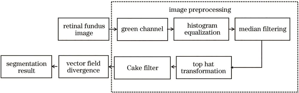
    Fig. 1. Block diagram of retinal blood vessel segmentation method based on multidirectional filtering
    Image segmentation results. (a) Retinal fundus image; (b) fundus image under the green channel; (c) image after histogram equalization and median filtering; (d) morphological top hat transformation image; (e) Cake filtered image; (f) segmentation result of proposed algorithm
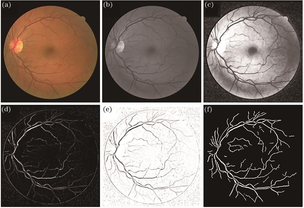
    Fig. 2. Image segmentation results. (a) Retinal fundus image; (b) fundus image under the green channel; (c) image after histogram equalization and median filtering; (d) morphological top hat transformation image; (e) Cake filtered image; (f) segmentation result of proposed algorithm
    Superimposed images in 6 directions. (a) Retinal fundus image; (b) extraction result at 0°; (c) extraction result at 30°; (d) extraction result at 60°; (e) extraction result at 90°; (f) extraction result at 120°; (g) extraction result at 150°; (h) segmentation result of proposed algorithm
    Fig. 3. Superimposed images in 6 directions. (a) Retinal fundus image; (b) extraction result at 0°; (c) extraction result at 30°; (d) extraction result at 60°; (e) extraction result at 90°; (f) extraction result at 120°; (g) extraction result at 150°; (h) segmentation result of proposed algorithm
    Segmentation results in the DRIVE data set. (a) Original fundus image; (b) standard image; (c) segmentation result of algorithm in Ref.[13]; (d) segmentation result of proposed algorithm
    Fig. 4. Segmentation results in the DRIVE data set. (a) Original fundus image; (b) standard image; (c) segmentation result of algorithm in Ref.[13]; (d) segmentation result of proposed algorithm
    Segmentation results in the STARE data set. (a) Original fundus image; (b) standard image; (c) segmentation result of algorithm in Ref.[13]; (d) segmentation result of proposed algorithm
    Fig. 5. Segmentation results in the STARE data set. (a) Original fundus image; (b) standard image; (c) segmentation result of algorithm in Ref.[13]; (d) segmentation result of proposed algorithm
    Partial view of blood vessel. (a) Original fundus partial image; (b) standard partial image; (c) segmentation result of algorithm in Ref.[13]; (d) segmentation result of proposed algorithm
    Fig. 6. Partial view of blood vessel. (a) Original fundus partial image; (b) standard partial image; (c) segmentation result of algorithm in Ref.[13]; (d) segmentation result of proposed algorithm
    AlgorithmDatabaseSensitivitySpecificityAccuracy
    Algorithm in Ref.[16DRIVE0.58110.93110.9383
    Algorithm in Ref.[17DRIVE0.72310.97590.9433
    Algorithm in Ref.[16STARE0.78650.96390.9581
    Proposed algorithmDRIVE0.98630.92670.9273
    Proposed algorithmSTARE0.92120.92850.9284
    Table 1. Performance of retinal vessel segmentation algorithms
    AlgorithmTime /sDatabase
    Algorithm in Ref.[1312.11DRIVE
    Proposed algorithm4.23DRIVE
    Algorithm in Ref.[138.75STARE
    Proposed algorithm5.67STARE
    Table 2. Running time of retinal vessel segmentation algorithms
    Caiyun Wang, Zhiyu Guan, Yida Wu, Chen Yao. Retinal Blood Vessel Segmentation Algorithm Based on Multidirectional Filtering[J]. Laser & Optoelectronics Progress, 2022, 59(8): 0817002
    Download Citation