
Bo Du, Xiang-Dong Chen, Ze-Hao Wang, Shao-Chun Zhang, En-Hui Wang, Guang-Can Guo, Fang-Wen Sun, "High resolution imaging with anomalous saturated excitation," Photonics Res. 9, 21 (2021)
- Photonics Research
- Vol. 9, Issue 1, 21 (2021)
![(a) Experimental setup. The pulsed 532 nm laser is circularly polarized, with a repetition rate of 5 MHz. DM: dichroic mirror; APD: avalanche photodiode. (b) The fluorescence emission of a single NV center with different laser intensities. The blue line is the fitting with Eq. (1). (c) Simulated point spread function and the spatial frequency distribution of the confocal microscopy with different powers. (d) The normalized spatial frequency with the excitation power of 0.06 and 1.2 mW. For comparison, we also show the results with a traditional saturating [b=0 in Eq. (1)]. The insert is the ratio of the high spatial frequency signal with different excitation laser powers.](/richHtml/prj/2021/9/1/01000021/img_001.jpg)
Fig. 1. (a) Experimental setup. The pulsed 532 nm laser is circularly polarized, with a repetition rate of 5 MHz. DM: dichroic mirror; APD: avalanche photodiode. (b) The fluorescence emission of a single NV center with different laser intensities. The blue line is the fitting with Eq. (1 ). (c) Simulated point spread function and the spatial frequency distribution of the confocal microscopy with different powers. (d) The normalized spatial frequency with the excitation power of 0.06 and 1.2 mW. For comparison, we also show the results with a traditional saturating [b = 0 1 )]. The insert is the ratio of the high spatial frequency signal with different excitation laser powers.
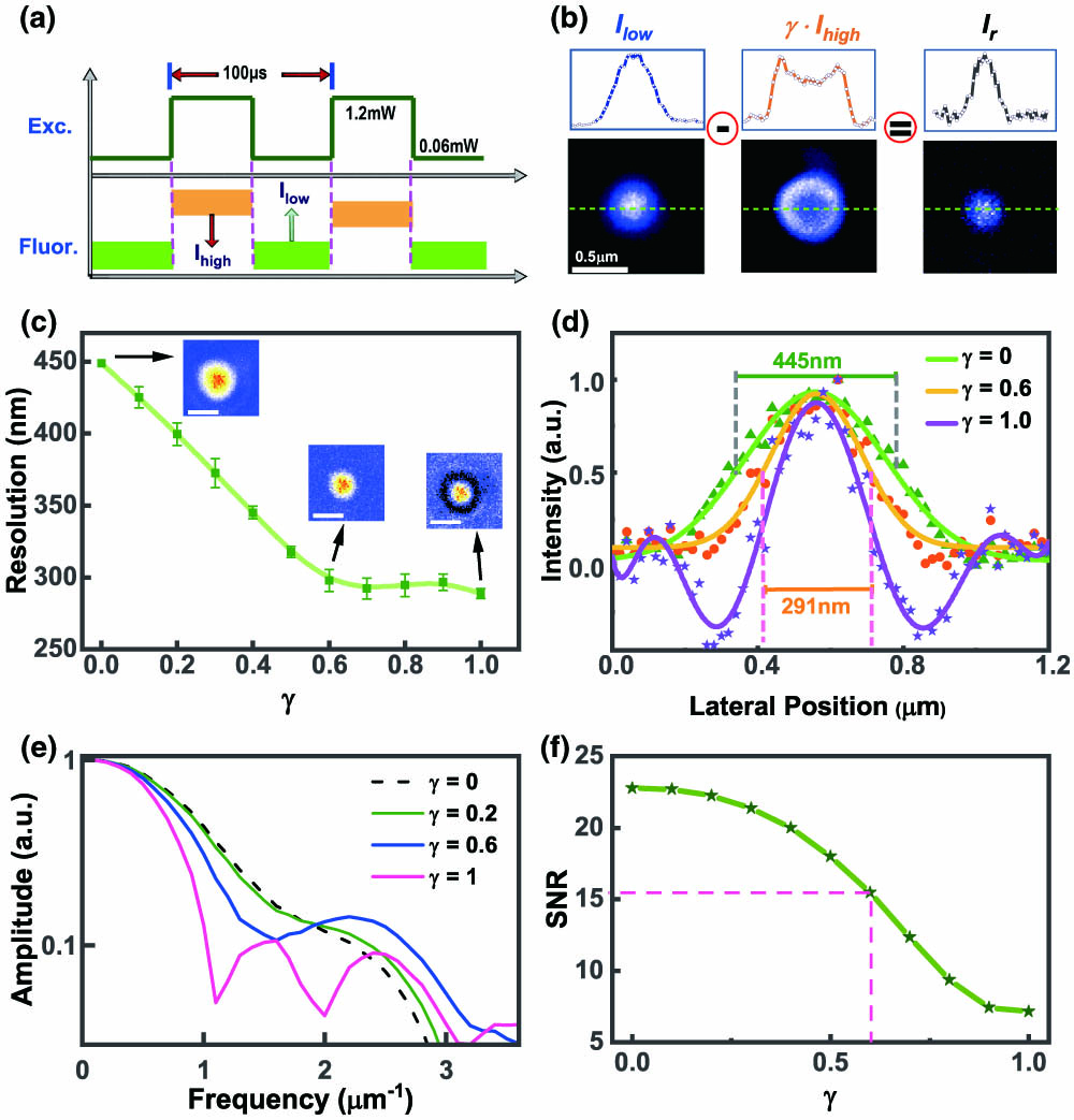
Fig. 2. (a) Sequences of the laser’s modulation and fluorescence detection. (b) The images of a single NV center. The confocal microscopy images of I low I high I r γ γ = 0 γ = 0 γ I r γ
Fig. 3. (a), (d) Multiple NV centers imaged with the confocal microscopies. (b), (e) Multiple NV centers imaged with ASAX microscopies. The scale bars are 1 μm in length. (c) and (f) are the cross-section profiles indicated by the arrows.


Set citation alerts for the article
Please enter your email address

© Copyright 2018-2021 | Chinese Laser Press. All Rights Reserved 沪ICP备15018463号-20