
Pengming Song, Shaowei Jiang, Tianbo Wang, Chengfei Guo, Ruihai Wang, Terrance Zhang, Guoan Zheng, "Synthetic aperture ptychography: coded sensor translation for joint spatial-Fourier bandwidth expansion," Photonics Res. 10, 1624 (2022)
- Photonics Research
- Vol. 10, Issue 7, 1624 (2022)
Abstract
1. INTRODUCTION
Ptychography is a coherent diffraction imaging (CDI) technique that has grown rapidly in the past years [1,2]. The original concept was developed to address the missing phase challenge in crystallography [3]. By translating a narrow coherent probe beam on a crystalline specimen, it aspires to extract the phase of Bragg peaks from far-field diffraction patterns. The modern form of this technique was brought to fruition by adopting an iterative phase retrieval framework [4]. Experiment procedures remain the same, where a specimen is laterally translated through a spatially confined probe beam and the diffraction patterns are recorded at the far field. Using the phase retrieval framework, the reconstruction process iteratively imposes two different constraints during the object scanning process: first, the diffraction measurements serve as the Fourier magnitude constraints in reciprocal space; second, the confined probe beam limits the physical extent of the object for each measurement and serves as the support constraint in real space. Ptychography requires no imaging optics downstream of a specimen under investigation. It inherently generates both intensity and phase contrast for quantitative investigation of material properties—a capability that many competing imaging modalities lack. The data redundancy of ptychography further allows it to recover other important system information, including the illumination probe beam [5–8], multiple coherent modes of the probe beam or the object [9], images at different spectral channels [10–12], multiple depth sections of a 3D object [13], diffraction data beyond the detector size [14], and orthogonal modes of an unstable illumination beam [15], among others. In the past decade, lensless ptychography has captured widespread interest from different imaging communities. In the field of biomedical imaging, it has been demonstrated for optofluidic flow-cytometer screening [16], urine sediment testing [17], blood analysis [17,18], malaria parasite screening [19], high-throughput digital pathology [19], antibiotic susceptibility testing [20], microbial limit testing [20], and large-scale yeast cell culture monitoring [21], among others. In the field of X-ray optics and extreme ultraviolet (EUV) imaging, it has become an indispensable modality in most synchrotrons and national laboratories worldwide [22,23]. Given that the brightness of coherent X-ray sources is expected to increase by orders of magnitude in the coming years, lensless ptychography has a promising future in imaging different non-crystalline structures on an atomic scale.
Ptychography can also be implemented using a lens-based system. Fourier ptychography (FP) is one example that swaps real space and reciprocal space using a lens [24]. In a typical FP implementation, an LED array is used to illuminate an object from different incident angles, and a low-numerical-aperture (NA) objective lens is used to acquire the corresponding low-resolution images. At each illumination angle, the captured image corresponds to the information of a circular aperture in Fourier space. The object scanning process in conventional ptychography is now implemented by the angular scanning process in FP for Fourier aperture synthesizing. The recovered spectrum in the reciprocal space of FP is then transformed back to the spatial domain to obtain a high-resolution, large-field-of-view object image with both intensity and phase properties. By combining with diffraction tomography, FP can also recover the scattering potential of 3D objects [25,26]. One surprising development of FP is a camera-scanning scheme that deviates from microscopy and enables far-field sub-diffraction imaging [27–29]. In this scheme, a lens–detector combo is translated at the far field for image acquisition. Far-field propagation of a coherent light field corresponds to the operation of a Fourier transform. Therefore, the entrance pupil of the lens naturally serves as a circular support constraint in reciprocal space. The lens then performs a second Fourier transform to convert the spectrum back to real space for imaging. By moving the entire lens–detector combo to different positions at the far field, one can acquire low-resolution images corresponding to different circular apertures in reciprocal space. These images can then be synthesized in a manner seen in FP. The size of the lens aperture does not limit the final image resolution. Instead, the resolution is determined by how far one can translate the camera.
In brief, conventional lensless ptychography scans an object in real space to widen the field of view. Lens-based FP scans the object spectrum in reciprocal space to expand the spatial-frequency bandwidth. These two modalities are mathematically equivalent if we treat the object spectrum in FP as the real-space object in conventional ptychography. Here we report an imaging scheme that merges the benefits of both techniques. This new scheme, termed synthetic aperture ptychography (SAP), is a lensless modality for far-field sub-diffraction imaging. Instead of translating the specimen over a confined probe beam, we illuminate the entire stationary object using an extended plane wave in SAP. A coded image sensor is then translated at the far field for
Sign up for Photonics Research TOC. Get the latest issue of Photonics Research delivered right to you!Sign up now
2. SYNTHETIC APERTURE PTYCHOGRAPHY
A. Concept and Operation of SAP
Drawing connections and distinctions within related modalities helps clarify the concept of the SAP approach. Figure 1 summarizes the differences between conventional ptychography and SAP. For both approaches, an object is placed in the spatial domain, and a detector is placed at the far field for diffraction data acquisition. When the detector is placed in the Fraunhofer zone with a Fresnel
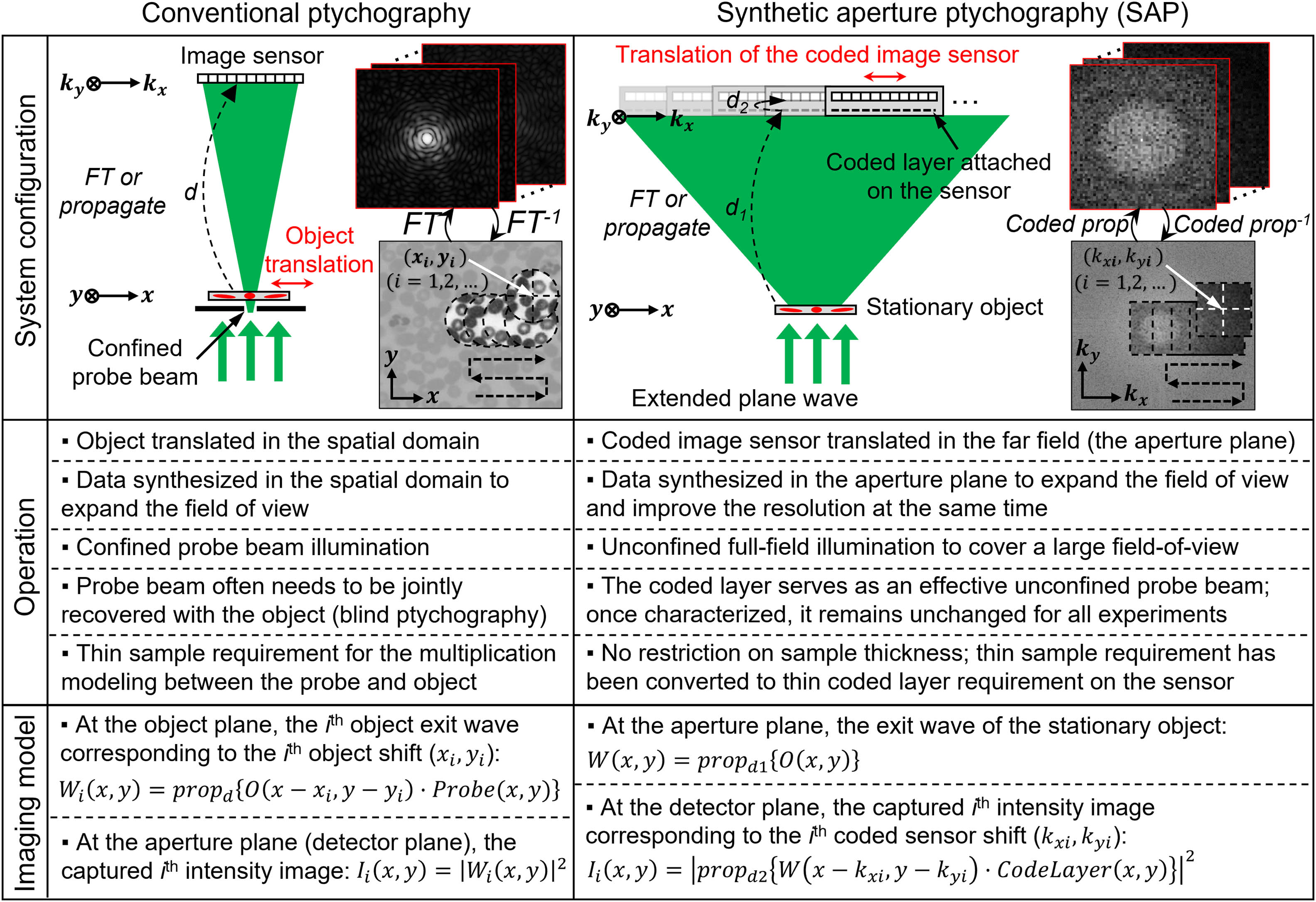
Figure 1.Comparison between conventional ptychography and SAP. Conventional ptychography translates the object over a confined probe beam in the spatial domain and acquires diffraction data at the far field. During the reconstruction process, conventional ptychography stitches the information in the spatial domain to expand the field of view. SAP illuminates the stationary object with an extended plane wave and translates a coded sensor at the far field for data acquisition. In the reconstruction process, SAP stitches the information in the intermediate aperture plane for object recovery. It can widen the field of view in real space and expand the spatial-frequency bandwidth in reciprocal space at the same time. The dashed arrows present free-space propagation over certain distances.
For conventional ptychography, the confined probe beam
For SAP, we illuminate the entire object with an extended plane wave. The exit waves from the object propagate for a distance
The advantages and unique properties of the SAP scheme can be summarized as follows. First, SAP stitches the information at the intermediate aperture plane when the detector is placed in the Fresnel zone. It can widen the imaging field of view and expand the Fourier bandwidth at the same time, getting the best of both conventional ptychography and FP. Second, the confined illumination probe beam in conventional ptychography often varies for different experiments. Therefore, the probe beam needs to be jointly recovered with the object in the reconstruction process (also termed blind ptychography [5–8]). In contrast, SAP adopts a fixed coded layer on the image sensor that, once characterized, remains unchanged for all subsequent experiments. Third, conventional ptychography uses a point-wise multiplication process to model the interaction between the object and the structured probe beam. This process is valid only when the sample is thin. The thickness limit for this approximation has been theoretically studied via different means [30,31]. In SAP, the object–probe multiplication process is performed at the coded layer plane. Therefore, the thin sample requirement in conventional ptychography is converted into a thin coded layer requirement in SAP [32], thus rendering the object thickness irrelevant to the reconstruction process. After recovery, one can digitally propagate the recovered object wavefront to any axial position for post-measurement refocusing [21,27,32]. Last, interferometric setups perform aperture synthesizing using a reference wave [33]. Such schemes require light sources with high spatial and temporal coherence. The associated optical setups are often subjected to vibration and other interferometry-related challenges. The reported SAP, on the other hand, requires neither interferometric measurements nor reference waves. It can be implemented with low-coherence sources such as X-ray and EUV sources, LEDs, and electrons. The lensless nature further makes it comparable to a variety of CDI setups that are generally limited by poor and aberrative lens elements.
B. Imaging Model and Reconstruction Process of SAP
In this section, we discuss the forward imaging model and the reconstruction process of SAP. As shown in the right panel of Fig. 1, the complex wavefront
Figure 2 shows the reconstruction process of SAP. We first initialize the object exit wavefront
![]()
Figure 2.Reconstruction process of SAP. The acquired images are used to update the specific regions of the exit wavefront
3. RESULTS
We validated the SAP approach with the object transmission and reflection configurations shown in Figs. 3(a) and 3(b). We illuminated the sample using a collimated 532-nm laser beam for both configurations. The coded mask on the image sensor was created by coating the sensor’s cover glass with microbeads. The size of the microbeads is 1–2 μm in diameter. This thin and dense coded layer allows the modulation process to be approximated by point-wise multiplication between the wavefront
![]()
Figure 3.Transmission and reflection configurations for SAP. For both configurations, the coded sensor is translated by a mechanical stage in the Fresnel zone. (a) Transmission configuration. The object exit wavefront synthesized at the intermediate aperture plane is backpropagated to the object plane for image recovery. (b) Reflection configuration. The coded sensor is translated in the direction perpendicular to the zero-order reflective beam. The recovered wavefront is then backpropagated to the object plane with a 20° tilted angle. (c) Employed coded image sensor. Two mask-free regions are used to track the positional shift in
For the transmission configuration in Fig. 3(a), we scanned the coded sensor in the Fresnel zone with
Figure 4 shows the experimental results of the transmission configuration. In this experiment, we used a resolution target as the object. Figure 4(a) shows the recovered complex object exit wavefront
Sign up for Photonics Research TOC. Get the latest issue of Photonics Research delivered right to you!Sign up now
![]()
Figure 4.Experimental validation of the transmission configuration of SAP. (a) Recovered wavefront
Figure 5 shows the experimental results of the reflection configuration. In this experiment, we used a microchip on a silicon wafer as the object. Figure 5(a) shows the recovered complex object exit wavefront at the coded layer plane. Similar to Fig. 4(a), the red dots in the left panels represent the scanning positions of the coded image sensor;
![]()
Figure 5.Experimental validation of the reflection configuration of SAP. (a) Recovered wavefront
Figures 4 and 5 validate the operation of the reported SAP approach. The sensor translation process in SAP synthesizes a large complex-valued wavefront at the intermediate aperture plane. For Figs. 4 and 5, we used eight iterations to recover the object exit wavefronts. By propagating this wavefront back to the object plane, we can widen the field of view in real space [Figs. 4(a) and 5(a)] and expand the spectral bandwidth in reciprocal space [Figs. 4(b) and 5(b)]. This unique feature enables us to obtain benefits from both conventional ptychography and FP. In Fig. S4 in Ref. [35], we compare the images at different planes in SAP. Figure S4(a) in Ref. [35] shows the raw images captured at the pixel-array plane. Figure S4(b) in Ref. [35] shows the synthesized images in the coded layer plane (intermediate aperture plane). Finally, Fig. S4(c) in Ref. [35] shows the images propagated back to the object plane.
A critical parameter for SAP reconstruction is the spatial overlap in between adjacent measurements. In Fig. S5, we analyze the SAP reconstructions with different spatial overlaps [35]. Figure S4(a) in Ref. [35] shows the SAP reconstructions using 400 raw measurements (
Another advantage of the reported SAP approach is its capability of imaging axially extended 3D objects. In the reconstruction process, we aim to recover the object exit wavefront
4. CONCLUSION
We report a new CDI modality termed SAP. In SAP, we illuminate the entire stationary object using an extended plane wave and translate a coded image sensor at the far field for data acquisition. When operated in the Fresnel zone, the translation of the coded sensor can effectively widen the imaging field of view and expand the NA for object recovery, thereby achieving a combination of benefits from both conventional ptychography and FP. The lensless nature of SAP further allows it to be implemented with a variety of CDI setups with radiation sources from visible light, EUV, and X-rays, to electrons. We also show that SAP waives the thin sample requirement in conventional ptychography. The exit wavefront recovered at the intermediate aperture plane can be backpropagated to different axial planes for post-acquisition refocusing.
We identify the following development areas for SAP. First, the development of better algorithms to recover or refine different system parameters is highly desired. In the current mPIE routine, the step size depends on a hyperparameter
The reported SAP offers a simple yet effective solution for sub-diffraction imaging via reference-free aperture synthesizing. The lensless nature of this modality further allows it to be implemented in a variety of CDIs that are generally limited by poor and aberrative lens elements. We envision that SAP will continue to grow and expand in various imaging applications.
Acknowledgment
Acknowledgment. P. S. acknowledges the support of the Thermo Fisher Scientific fellowship.
References
[1] J. Rodenburg, A. Maiden. Ptychography. Springer Handbook of Microscopy, 819-904(2019).
[2] M. Guizar-Sicairos, P. Thibault. Ptychography: a solution to the phase problem. Phys. Today, 74, 42-48(2021).
[3] W. Hoppe, G. Strube. Diffraction in inhomogeneous primary wave fields. 2. Optical experiments for phase determination of lattice interferences. Acta Crystallogr. A, 25, 502-507(1969).
[4] H. M. L. Faulkner, J. Rodenburg. Movable aperture lensless transmission microscopy: a novel phase retrieval algorithm. Phys. Rev. Lett., 93, 023903(2004).
[5] M. Guizar-Sicairos, J. R. Fienup. Phase retrieval with transverse translation diversity: a nonlinear optimization approach. Opt. Express, 16, 7264-7278(2008).
[6] A. M. Maiden, J. M. Rodenburg. An improved ptychographical phase retrieval algorithm for diffractive imaging. Ultramicroscopy, 109, 1256-1262(2009).
[7] P. Thibault, M. Dierolf, O. Bunk, A. Menzel, F. Pfeiffer. Probe retrieval in ptychographic coherent diffractive imaging. Ultramicroscopy, 109, 338-343(2009).
[8] X. Ou, G. Zheng, C. Yang. Embedded pupil function recovery for Fourier ptychographic microscopy. Opt. Express, 22, 4960-4972(2014).
[9] P. Thibault, A. Menzel. Reconstructing state mixtures from diffraction measurements. Nature, 494, 68-71(2013).
[10] D. J. Batey, D. Claus, J. M. Rodenburg. Information multiplexing in ptychography. Ultramicroscopy, 138, 13-21(2014).
[11] P. Song, R. Wang, J. Zhu, T. Wang, Z. Bian, Z. Zhang, K. Hoshino, M. Murphy, S. Jiang, C. Guo. Super-resolved multispectral lensless microscopy via angle-tilted, wavelength-multiplexed ptychographic modulation. Opt. Lett., 45, 3486-3489(2020).
[12] Y. Yao, Y. Jiang, J. Klug, Y. Nashed, C. Roehrig, C. Preissner, F. Marin, M. Wojcik, O. Cossairt, Z. Cai. Broadband X-ray ptychography using multi-wavelength algorithm. J. Synchrotron. Radiat., 28, 309-317(2021).
[13] A. M. Maiden, M. J. Humphry, J. Rodenburg. Ptychographic transmission microscopy in three dimensions using a multi-slice approach. J. Opt. Soc. Am. A, 29, 1606-1614(2012).
[14] A. M. Maiden, M. J. Humphry, F. Zhang, J. M. Rodenburg. Superresolution imaging via ptychography. J. Opt. Soc. Am. A, 28, 604-612(2011).
[15] M. Odstrcil, P. Baksh, S. Boden, R. Card, J. Chad, J. Frey, W. Brocklesby. Ptychographic coherent diffractive imaging with orthogonal probe relaxation. Opt. Express, 24, 8360-8369(2016).
[16] P. Song, C. Guo, S. Jiang, T. Wang, P. Hu, D. Hu, Z. Zhang, B. Feng, G. Zheng. Optofluidic ptychography on a chip. Lab Chip, 21, 4549-4556(2021).
[17] S. Jiang, C. Guo, T. Wang, J. Liu, P. Song, T. Zhang, R. Wang, B. Feng, G. Zheng. Blood-coated sensor for high-throughput ptychographic cytometry on a Blu-Ray disc. ACS Sens., 7, 1058-1067(2022).
[18] S. Jiang, C. Guo, P. Song, N. Zhou, Z. Bian, J. Zhu, R. Wang, P. Dong, Z. Zhang, J. Liao, J. Yao, B. Feng, M. Murphy, G. Zheng. Resolution-enhanced parallel coded ptychography for high-throughput optical imaging. ACS Photon., 8, 3261-3271(2021).
[19] S. Jiang, C. Guo, P. Song, T. Wang, R. Wang, T. Zhang, Q. Wu, R. Pandey, G. Zheng. High-throughput digital pathology via a handheld, multiplexed, and AI-powered ptychographic whole slide scanner. Lab Chip(2022).
[20] S. Jiang, C. Guo, Z. Bian, R. Wang, J. Zhu, P. Song, P. Hu, D. Hu, Z. Zhang, K. Hoshino, B. Feng, G. Zheng. Ptychographic sensor for large-scale lensless microbial monitoring with high spatiotemporal resolution. Biosens. Bioelectron., 196, 113699(2022).
[21] S. Jiang, J. Zhu, P. Song, C. Guo, Z. Bian, R. Wang, Y. Huang, S. Wang, H. Zhang, G. Zheng. Wide-field, high-resolution lensless on-chip microscopy via near-field blind ptychographic modulation. Lab Chip, 20, 1058-1065(2020).
[22] L. Loetgering, S. Witte, J. Rothhardt. Advances in laboratory-scale ptychography using high harmonic sources. Opt. Express, 30, 4133-4164(2022).
[23] F. Pfeiffer. X-ray ptychography. Nat. Photonics, 12, 9-17(2018).
[24] G. Zheng, R. Horstmeyer, C. Yang. Wide-field, high-resolution Fourier ptychographic microscopy. Nat. Photonics, 7, 739-745(2013).
[25] R. Horstmeyer, J. Chung, X. Ou, G. Zheng, C. Yang. Diffraction tomography with Fourier ptychography. Optica, 3, 827-835(2016).
[26] C. Zuo, J. Sun, J. Li, A. Asundi, Q. Chen. Wide-field high-resolution 3D microscopy with Fourier ptychographic diffraction tomography. Opt. Lasers Eng., 128, 106003(2020).
[27] S. Dong, R. Horstmeyer, R. Shiradkar, K. Guo, X. Ou, Z. Bian, H. Xin, G. Zheng. Aperture-scanning Fourier ptychography for 3D refocusing and super-resolution macroscopic imaging. Opt. Express, 22, 13586-13599(2014).
[28] J. Holloway, Y. Wu, M. K. Sharma, O. Cossairt, A. Veeraraghavan. SAVI: synthetic apertures for long-range, subdiffraction-limited visible imaging using Fourier ptychography. Sci. Adv., 3, e1602564(2017).
[29] K. Wakonig, A. Diaz, A. Bonnin, M. Stampanoni, A. Bergamaschi, J. Ihli, M. Guizar-Sicairos, A. Menzel. X-ray Fourier ptychography. Sci. Adv., 5, eaav0282(2019).
[30] P. Thibault, M. Dierolf, A. Menzel, O. Bunk, C. David, F. Pfeiffer. High-resolution scanning x-ray diffraction microscopy. Science, 321, 379-382(2008).
[31] E. H. Tsai, I. Usov, A. Diaz, A. Menzel, M. Guizar-Sicairos. X-ray ptychography with extended depth of field. Opt. Express, 24, 29089-29108(2016).
[32] P. Song, S. Jiang, H. Zhang, Z. Bian, C. Guo, K. Hoshino, G. Zheng. Super-resolution microscopy via ptychographic structured modulation of a diffuser. Opt. Lett., 44, 3645-3648(2019).
[33] A. E. Tippie, A. Kumar, J. R. Fienup. High-resolution synthetic-aperture digital holography with digital phase and pupil correction. Opt. Express, 19, 12027-12038(2011).
[34] A. Maiden, D. Johnson, P. Li. Further improvements to the ptychographical iterative engine. Optica, 4, 736-745(2017).
[36] S. Jiang, C. Guo, P. Hu, D. Hu, P. Song, T. Wang, Z. Bian, Z. Zhang, G. Zheng. High-throughput lensless whole slide imaging via continuous height-varying modulation of a tilted sensor. Opt. Lett., 46, 5212-5215(2021).
[37] G. Zheng, C. Kolner, C. Yang. Microscopy refocusing and dark-field imaging by using a simple LED array. Opt. Lett., 36, 3987-3989(2011).
[38] M. Odstrčil, A. Menzel, M. Guizar-Sicairos. Iterative least-squares solver for generalized maximum-likelihood ptychography. Opt. Express, 26, 3108-3123(2018).


Set citation alerts for the article
Please enter your email address

© Copyright 2018-2021 | Chinese Laser Press. All Rights Reserved 沪ICP备15018463号-20