• Chinese Optics Letters
  • Vol. 16, Issue 8, 081701 (2018)
Zhongwen Cheng1、2, Haigang Ma2, Zhiyang Wang2, and Sihua Yang1、2、*
Author Affiliations
  • 1MOE Key Laboratory of Laser Life Science & Institute of Laser Life Science, South China Normal University, Guangzhou 510631, China
  • 2College of Biophotonics, South China Normal University, Guangzhou 510631, China
  • show less
    DOI: 10.3788/COL201816.081701 Cite this Article Set citation alerts
    Zhongwen Cheng, Haigang Ma, Zhiyang Wang, Sihua Yang. 3D depth-coded photoacoustic microscopy with a large field of view for human skin imaging[J]. Chinese Optics Letters, 2018, 16(8): 081701 Copy Citation Text show less
    Schematic of the 3D depth-coded PA microscopy. (a) Schematic of the imaging system. (b) A photo of a large-field mechanical scanning head. (c) The structure of the internal core component in (b). SMF, single-mode fiber; NDF, neutral density filter; PD, photodiode; DAQ, data acquisition; AL, aspheric lens; UT, ultrasonic transducer; FPGA, field programmable gate array.
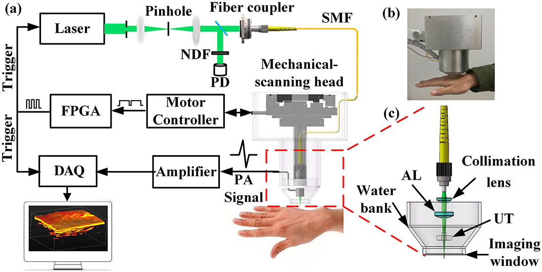
    Fig. 1. Schematic of the 3D depth-coded PA microscopy. (a) Schematic of the imaging system. (b) A photo of a large-field mechanical scanning head. (c) The structure of the internal core component in (b). SMF, single-mode fiber; NDF, neutral density filter; PD, photodiode; DAQ, data acquisition; AL, aspheric lens; UT, ultrasonic transducer; FPGA, field programmable gate array.
    Resolution of the PA microscopy. (a) Pulse response of the ultrasonic transducer at the focus. (b) Amplitude frequency response of the ultrasonic transducer. (c) The lateral resolution of the PA microscopy. (d) The axial resolution of the PA microscopy.
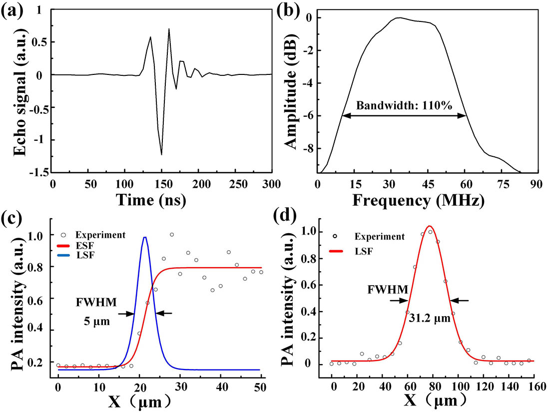
    Fig. 2. Resolution of the PA microscopy. (a) Pulse response of the ultrasonic transducer at the focus. (b) Amplitude frequency response of the ultrasonic transducer. (c) The lateral resolution of the PA microscopy. (d) The axial resolution of the PA microscopy.
    PA images of a leaf vein. (a) Schematic illustration of the experiment. (b) MIP PA image of the leaf vein. (c) Volumetric PA image of the leaf vein. (d) 3D depth-coded PA image corresponding to (c).
    Fig. 3. PA images of a leaf vein. (a) Schematic illustration of the experiment. (b) MIP PA image of the leaf vein. (c) Volumetric PA image of the leaf vein. (d) 3D depth-coded PA image corresponding to (c).
    PA images of the rabbit ear. (a) Photos of the rabbit ear as well as the imaging area. (b) The PA volumetric vascular image. (c) The MIP PA image. (d) The 3D depth-coded PA image.
    Fig. 4. PA images of the rabbit ear. (a) Photos of the rabbit ear as well as the imaging area. (b) The PA volumetric vascular image. (c) The MIP PA image. (d) The 3D depth-coded PA image.
    PA images of human opisthenar and palm. (a), (b) Photos of the human opisthenar and palm, respectively. The insets show the corresponding imaging area. (c), (d) PA volumetric images of the opisthenar (Media 1) and palm (Media 2), respectively. (e), (f) PA volumetric vascular images of the opisthenar and palm beneath the epidermis seen from top to bottom. (g), (h) PA volumetric vascular images of the opisthenar and palm beneath the epidermis seen from bottom to top. (i), (j) The 3D depth-coded PA images of the opisthenar and palm corresponding to (e) and (f).
    Fig. 5. PA images of human opisthenar and palm. (a), (b) Photos of the human opisthenar and palm, respectively. The insets show the corresponding imaging area. (c), (d) PA volumetric images of the opisthenar (Media 1) and palm (Media 2), respectively. (e), (f) PA volumetric vascular images of the opisthenar and palm beneath the epidermis seen from top to bottom. (g), (h) PA volumetric vascular images of the opisthenar and palm beneath the epidermis seen from bottom to top. (i), (j) The 3D depth-coded PA images of the opisthenar and palm corresponding to (e) and (f).
    Zhongwen Cheng, Haigang Ma, Zhiyang Wang, Sihua Yang. 3D depth-coded photoacoustic microscopy with a large field of view for human skin imaging[J]. Chinese Optics Letters, 2018, 16(8): 081701
    Download Citation