• Photonics Research
  • Vol. 6, Issue 4, 332 (2018)
Yuchen Wang1、2, Shu Hu1, Xiao Yang3, Ruizhi Wang1, Heng Li1, and Chuanxiang Sheng1、*
Author Affiliations
  • 1School of Electronic and Optical Engineering, Nanjing University of Science and Technology, Nanjing 210094, China
  • 2School of Science, Qingdao University of Technology, Qingdao 266520, China
  • 3Faculty of Electronic Information Engineering, Huaiyin Institute of Technology, Huaian 223003, China
  • show less
    DOI: 10.1364/PRJ.6.000332 Cite this Article Set citation alerts
    Yuchen Wang, Shu Hu, Xiao Yang, Ruizhi Wang, Heng Li, Chuanxiang Sheng. Evanescent-wave pumped single-mode microcavity laser from fiber of 125  μm diameter[J]. Photonics Research, 2018, 6(4): 332 Copy Citation Text show less
    (a) Diagram of the experimental setup; (b) comparison between photoluminescence (PL) spectrum excited by a continuous wave laser at 20 mW/cm2 and the amplified spontaneous emission (ASE) excited by a 10 ns pulsed laser at 96.5 μJ/pulse of 5 mg/mL ethanol solution of rhodamine 6G (Rh6G). Inset is the molecular structure of Rh6G.
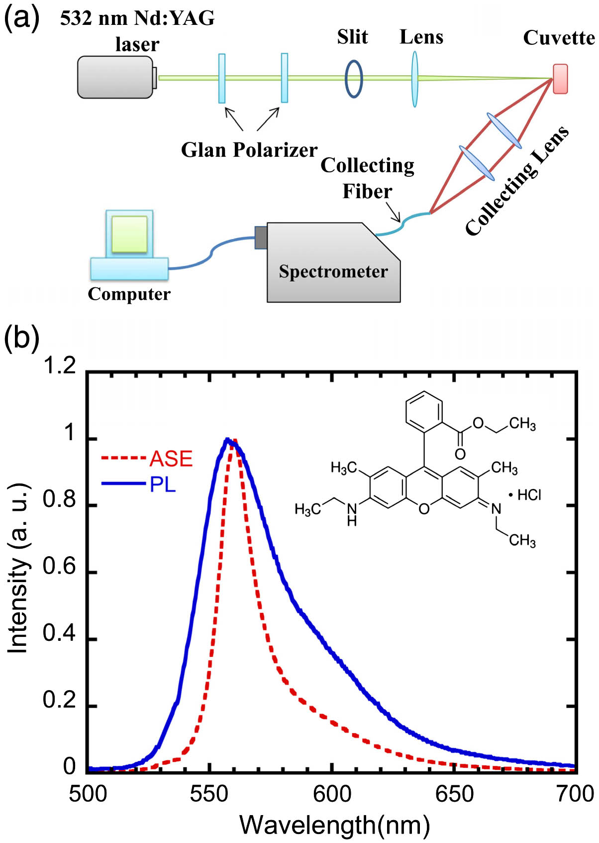
    Fig. 1. (a) Diagram of the experimental setup; (b) comparison between photoluminescence (PL) spectrum excited by a continuous wave laser at 20  mW/cm2 and the amplified spontaneous emission (ASE) excited by a 10 ns pulsed laser at 96.5 μJ/pulse of 5 mg/mL ethanol solution of rhodamine 6G (Rh6G). Inset is the molecular structure of Rh6G.
    (a) Emission spectra of a 5 mg/mL Rh6G ethanol solution with a fiber at various excitation intensities. Left inset: sample configuration; the bare fiber rests on the cuvette wall vertically by capillary force. Right inset: emission spectrum excited at 3.7 μJ/pulse. TE, transverse electric; TM, transverse magnetic. (b) Integrated intensity of the emission as a function of excitation pulse energy, indicating a threshold behavior.
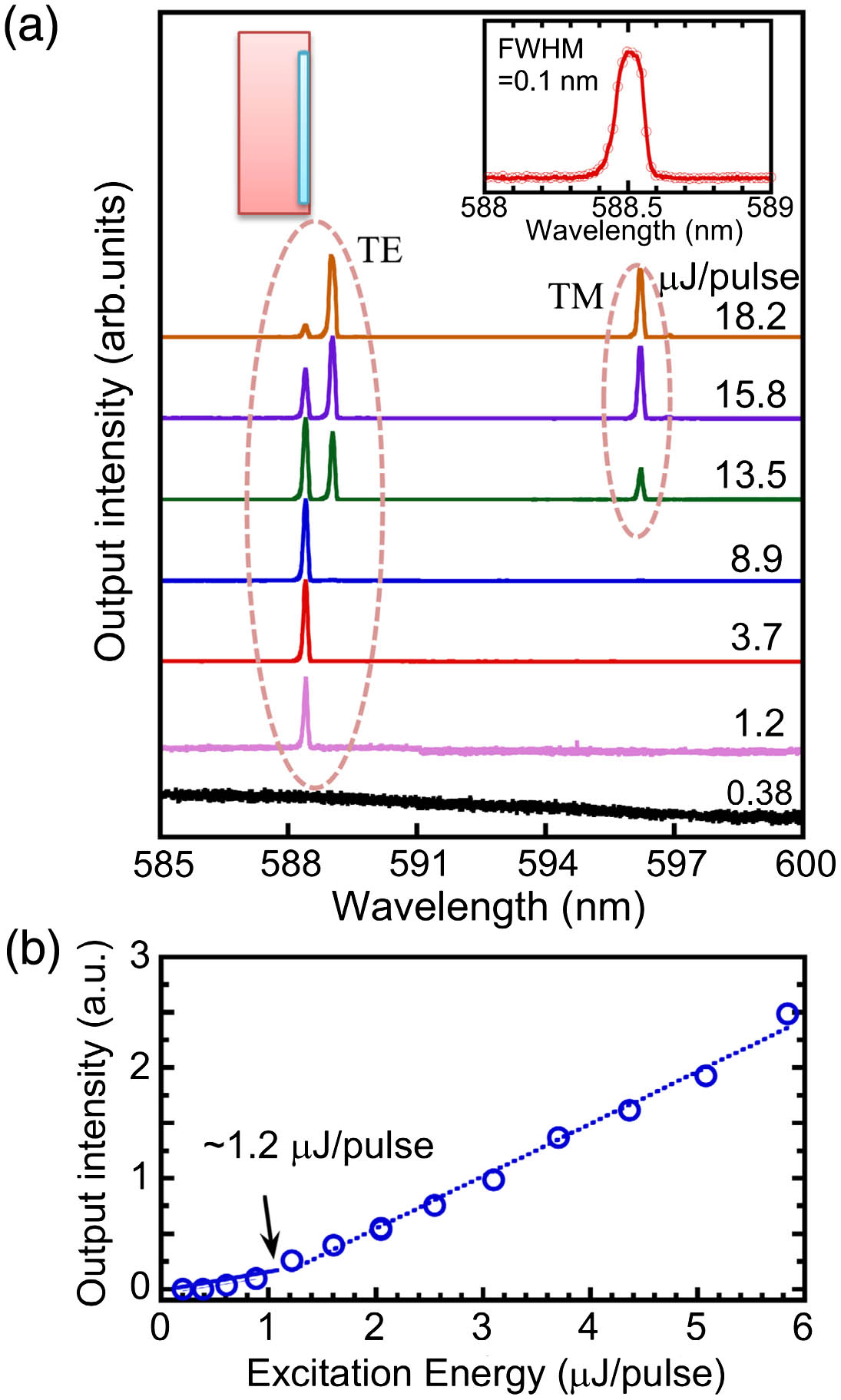
    Fig. 2. (a) Emission spectra of a 5 mg/mL Rh6G ethanol solution with a fiber at various excitation intensities. Left inset: sample configuration; the bare fiber rests on the cuvette wall vertically by capillary force. Right inset: emission spectrum excited at 3.7 μJ/pulse. TE, transverse electric; TM, transverse magnetic. (b) Integrated intensity of the emission as a function of excitation pulse energy, indicating a threshold behavior.
    Single-mode emission from various samples and from various excitation positions in the same sample, all excited using similar pulse energy (∼10 μJ/pulse). S1 (2, 3): sample 1 (2, 3). S1. P1(2, 3): position 1 (2, 3) in sample 1.
    Fig. 3. Single-mode emission from various samples and from various excitation positions in the same sample, all excited using similar pulse energy (10  μJ/pulse). S1 (2, 3): sample 1 (2, 3). S1. P1(2, 3): position 1 (2, 3) in sample 1.
    (a) Scheme of a fiber with coating layer at one end in the 5 mg/mL Rh6G ethanol solution. Two positions on the fiber are marked as 1, 2, respectively; (b) corresponding spectrum excited at two positions by a nanosecond (ns) pulsed laser at pump energy of ∼8 μJ/pulse.
    Fig. 4. (a) Scheme of a fiber with coating layer at one end in the 5 mg/mL Rh6G ethanol solution. Two positions on the fiber are marked as 1, 2, respectively; (b) corresponding spectrum excited at two positions by a nanosecond (ns) pulsed laser at pump energy of 8  μJ/pulse.
    (a) WGM spectra at various excitation intensities from a fiber in a 5 mg/mL Rh6G ethanol solution. Sample configuration was schematically shown in the left inset, with a bare fiber being coated with PMMA (∼thickness of 1 μm) at both ends. Right inset: typical emission spectrum of one WGM mode with FWHM of 0.1 nm; (b) integrated intensity of the emission as a function of excitation pulse energy, indicating a threshold behavior.
    Fig. 5. (a) WGM spectra at various excitation intensities from a fiber in a 5 mg/mL Rh6G ethanol solution. Sample configuration was schematically shown in the left inset, with a bare fiber being coated with PMMA (thickness of 1 μm) at both ends. Right inset: typical emission spectrum of one WGM mode with FWHM of 0.1 nm; (b) integrated intensity of the emission as a function of excitation pulse energy, indicating a threshold behavior.
    WGM spectra of a fiber in 5 mg/mL Rh6G solution of mixed ethanol and ethylene glycol with different ratios. The pump energy is ∼10 μJ/pulse; in this configuration, the fiber sticks to the cuvette wall. The corresponding refractive indexes are marked at each spectrum. The single-mode emission spectra, which are circled, are also zoomed to show the lack of spectral shift (left inset). Right inset is the WGM emission from a bare fiber in MEH-PPV/THF solution using the same configuration.
    Fig. 6. WGM spectra of a fiber in 5 mg/mL Rh6G solution of mixed ethanol and ethylene glycol with different ratios. The pump energy is 10  μJ/pulse; in this configuration, the fiber sticks to the cuvette wall. The corresponding refractive indexes are marked at each spectrum. The single-mode emission spectra, which are circled, are also zoomed to show the lack of spectral shift (left inset). Right inset is the WGM emission from a bare fiber in MEH-PPV/THF solution using the same configuration.
    Plot of γ(λ), which is the fraction of the excited molecules at the laser threshold for various Qsolution=ηQ; Q is the quality factor of the cavity, and η is the occupation factor, which is defined as the fraction of the evanescent field volume to that of the whole WGM [27].
    Fig. 7. Plot of γ(λ), which is the fraction of the excited molecules at the laser threshold for various Qsolution=ηQ; Q is the quality factor of the cavity, and η is the occupation factor, which is defined as the fraction of the evanescent field volume to that of the whole WGM [27].
    Emission spectra from a fiber in 5 mg/mL rhodamine B ethanol solution at various excitation intensities with the same sample configuration as in Fig. 2(a). Left inset: PL spectrum of the solution excited at 20 mW/cm2; right inset: molecular structure of rhodamine B.
    Fig. 8. Emission spectra from a fiber in 5 mg/mL rhodamine B ethanol solution at various excitation intensities with the same sample configuration as in Fig. 2(a). Left inset: PL spectrum of the solution excited at 20  mW/cm2; right inset: molecular structure of rhodamine B.
    Yuchen Wang, Shu Hu, Xiao Yang, Ruizhi Wang, Heng Li, Chuanxiang Sheng. Evanescent-wave pumped single-mode microcavity laser from fiber of 125  μm diameter[J]. Photonics Research, 2018, 6(4): 332
    Download Citation