• Opto-Electronic Engineering
  • Vol. 49, Issue 3, 210361-1 (2022)
Ruqian Hao, Xiangzhou Wang, Jing Zhang, Juanxiu Liu*, Xiaohui Du, and Lin Liu
Author Affiliations
  • School of Optoelectronic Science and Engineering, University of Electronic Science and Technology of China, Chengdu, Sichuan 611731, China
  • show less
    DOI: 10.12086/oee.2022.210361 Cite this Article
    Ruqian Hao, Xiangzhou Wang, Jing Zhang, Juanxiu Liu, Xiaohui Du, Lin Liu. An automatic object detection method for microscopic images based on attention mechanism[J]. Opto-Electronic Engineering, 2022, 49(3): 210361-1 Copy Citation Text show less
    The basic blocks of ResNet and ResNeXt. (a) Basic block of ResNet; (b) Basic block of ResNeXt
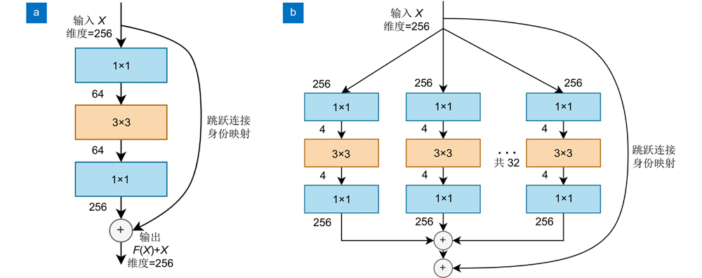
    Fig. 1. The basic blocks of ResNet and ResNeXt. (a) Basic block of ResNet; (b) Basic block of ResNeXt
    Workflow of the proposed algorithm
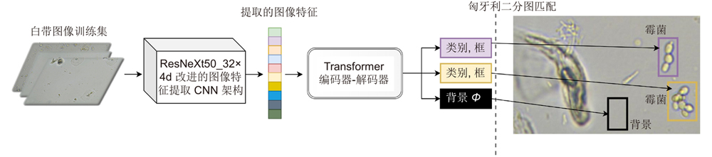
    Fig. 2. Workflow of the proposed algorithm
    Microscopic images of the three common pathogenic cells that cause vaginitis. (a) Mildew; (b) Trichomonas; (c) Clue cell
    Fig. 3. Microscopic images of the three common pathogenic cells that cause vaginitis. (a) Mildew; (b) Trichomonas; (c) Clue cell
    The performance of the original DETR and the improved DETR on validation dataset
    Fig. 4. The performance of the original DETR and the improved DETR on validation dataset
    The comparison of PR curves computed from the original model and the improved model. (a) PR curve of mAP; (b) PR curve of mildew; (c) PR curve of trichomonas; (d) PR curve of clue cell
    Fig. 5. The comparison of PR curves computed from the original model and the improved model. (a) PR curve of mAP; (b) PR curve of mildew; (c) PR curve of trichomonas; (d) PR curve of clue cell
    The detection results of the three common pathogenic cells. (a) Detection results of mildew; (b) Detection results of trichomonas; (c) Detection results of clue cell
    Fig. 6. The detection results of the three common pathogenic cells. (a) Detection results of mildew; (b) Detection results of trichomonas; (c) Detection results of clue cell
    Comparison of detection results and attention weights visualization map. (a) Original image; (b) Ground truth; (c) Detection results of original DETR; (d) Attention weights visualization of the original DETR; (e) Attention weights visualization of the original DETR on the original image; (f) Detection results of the improved DETR; (g) Attention weights visualization of the improved DETR; (h) Attention weights visualization of the improved DETR on the original image
    Fig. 7. Comparison of detection results and attention weights visualization map. (a) Original image; (b) Ground truth; (c) Detection results of original DETR; (d) Attention weights visualization of the original DETR; (e) Attention weights visualization of the original DETR on the original image; (f) Detection results of the improved DETR; (g) Attention weights visualization of the improved DETR; (h) Attention weights visualization of the improved DETR on the original image
    数据集类别图像张数标注细胞总数霉菌个数滴虫个数线索细胞个数
    训练集653184417576324
    验证集2185935622110
    测试集218620582308
    Table 1. The details of dataset split
    模型mAP/%霉菌AP/%滴虫AP/%线索细胞AP/%
    原始DETR87.587.085.989.7
    本文改进模型96.393.095.8100.0
    Table 2. The comparison of mAP and AP results of the original model and the proposed model
    评价指标模型霉菌滴虫线索细胞
    查准率(precision)/%原始DETR85.390.0100
    本文改进模型88.0100100
    召回率(recall)/%原始DETR85.090.075.0
    本文改进模型91.093.0100
    Table 3. The comparison of precision and recall results of the original DETR and the proposed improved DETR
    Ruqian Hao, Xiangzhou Wang, Jing Zhang, Juanxiu Liu, Xiaohui Du, Lin Liu. An automatic object detection method for microscopic images based on attention mechanism[J]. Opto-Electronic Engineering, 2022, 49(3): 210361-1
    Download Citation