• Spectroscopy and Spectral Analysis
  • Vol. 42, Issue 7, 2200 (2022)
Qi-yan ZHANG1、*, Xiao LIU1、*, Jie YANG2、2; 3; *;, Wei-xin SHI1、1;, Qing-nan GAO1、1;, Hong ZHANG1、1;, and Huang DENG1、1;
Author Affiliations
  • 11. Core and Samples Center of Land and Resources, China Geological Survey, Beijing 100083, China
  • 22. State Key Laboratory of Geological Process and Mineral Resources, China University of Geosciences (Beijing), Beijing 100083, China
  • show less
    DOI: 10.3964/j.issn.1000-0593(2022)07-2200-07 Cite this Article
    Qi-yan ZHANG, Xiao LIU, Jie YANG, Wei-xin SHI, Qing-nan GAO, Hong ZHANG, Huang DENG. Application of Micro X-Ray Fluorescence Imaging Technology in Core Analysis[J]. Spectroscopy and Spectral Analysis, 2022, 42(7): 2200 Copy Citation Text show less
    The maps of element disribution
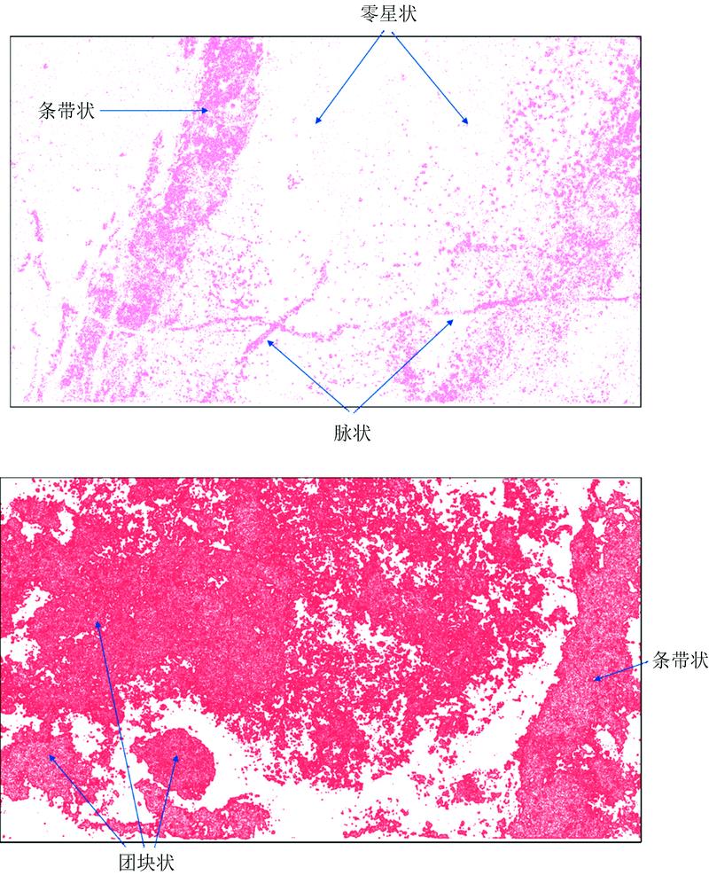
    Fig. 1. The maps of element disribution
    Element distribution map of copper-bearing hornstones with skarn breccia(a): Photo of sample; (b)—(i): Elemental maps using micro X-Ray Fluorescence
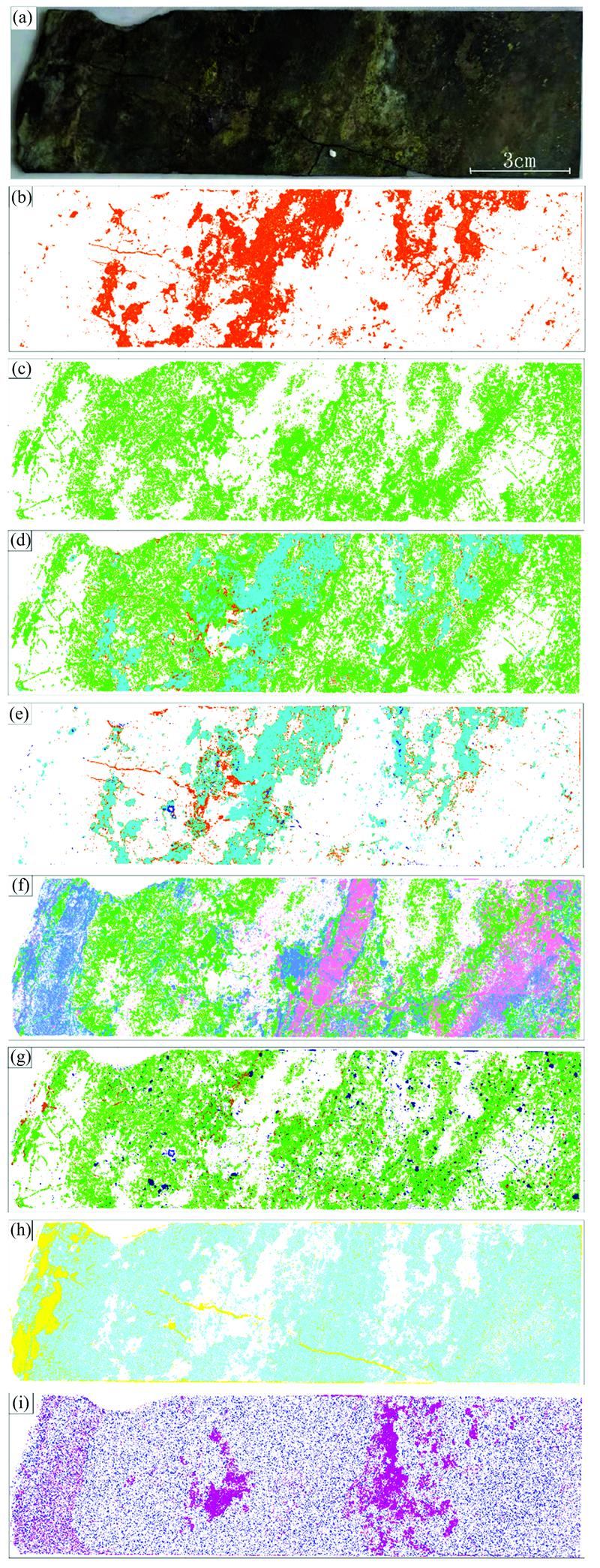
    Fig. 2. Element distribution map of copper-bearing hornstones with skarn breccia
    (a): Photo of sample; (b)—(i): Elemental maps using micro X-Ray Fluorescence
    Element distribution map of copper-bearing siliceous rock(a): Photo of sample; (b)—(i): Elemental maps using micro X-Ray Fluorescence
    Fig. 3. Element distribution map of copper-bearing siliceous rock
    (a): Photo of sample; (b)—(i): Elemental maps using micro X-Ray Fluorescence
    Element distribution map of copper-bearing skarn(a): Photo of sample; (b)—(j): Elemental maps using micro X-Ray Fluorescence
    Fig. 4. Element distribution map of copper-bearing skarn
    (a): Photo of sample; (b)—(j): Elemental maps using micro X-Ray Fluorescence
    Element distribution map of copper-bearing pyrrhotite ore(a): Photo of sample; (b)—(i): Elemental maps using micro X-Ray Fluorescence
    Fig. 5. Element distribution map of copper-bearing pyrrhotite ore
    (a): Photo of sample; (b)—(i): Elemental maps using micro X-Ray Fluorescence
    样品名起始深度/m终止深度/m样品尺寸/cm地层主要矿石矿物主要脉石矿物
    含铜角岩夹矽卡岩492.23492.4017×5.0三叠系下统小凉亭组黄铜矿、 黄铁矿石英、 石榴子石、 透辉石、 绿泥石、 方解石
    含铜硅质岩852.68852.9022×4.5二叠系下统栖霞组黄铜矿石英、 石榴子石、 透辉石
    含铜矽卡岩1 124.941 125.1016×4.6石炭系中、 上统黄龙、 船山组黄铜矿、 黄铁矿、 磁黄铁矿石榴子石、 透辉石、 石英、 绿泥石
    含铜磁黄铁矿石1 156.701 156.9020×4.7石炭系中、 上统黄龙、 船山组磁黄铁矿、 黄铜矿、 磁铁矿绿泥石、 透闪石
    Table 1. Geological information of core samples
    样品名宽/
    pixel
    高/
    pixel
    总像素/pixel单像素大小/
    μm
    停留时间/
    (ms·pixel-1)
    移动速度/
    (mm·s-1)
    含铜角岩夹矽卡岩3 4809983473 04050105
    含铜硅质岩4 5459014 095 0455086.3
    含铜矽卡岩3 3989323 166 9365086.3
    含铜磁黄铁矿石4 2459373 977 56550105
    Table 2. Scan parameters of Micro-XRF
    Qi-yan ZHANG, Xiao LIU, Jie YANG, Wei-xin SHI, Qing-nan GAO, Hong ZHANG, Huang DENG. Application of Micro X-Ray Fluorescence Imaging Technology in Core Analysis[J]. Spectroscopy and Spectral Analysis, 2022, 42(7): 2200
    Download Citation