• Chinese Optics Letters
  • Vol. 14, Issue 11, 111102 (2016)
Jian Zhang1, Jiadong Fan1, Jianhua Zhang1, Qingjie Huang2, and Huaidong Jiang1、3、*
Author Affiliations
  • 1State Key Laboratory of Crystal Materials, Shandong University, Jinan 250100, China
  • 2School of Information Science and Engineering, Shandong University, Jinan 250100, China
  • 3School of Physical Science and Technology, ShanghaiTech University, Shanghai 201210, China
  • show less
    DOI: 10.3788/COL201614.111102 Cite this Article Set citation alerts
    Jian Zhang, Jiadong Fan, Jianhua Zhang, Qingjie Huang, Huaidong Jiang. 3D imaging by two-color Ewald spheres with optical lasers[J]. Chinese Optics Letters, 2016, 14(11): 111102 Copy Citation Text show less
    Schematic diagram of a coherent diffraction microscope with two lasers at 543 (green) and 432 nm (blue) in the same orientation.
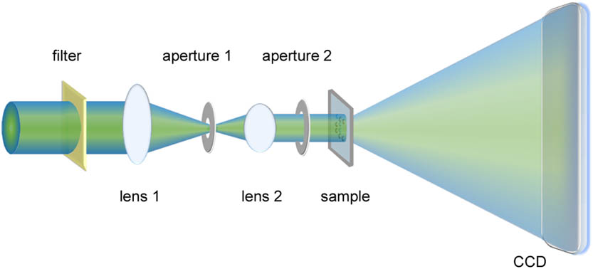
    Fig. 1. Schematic diagram of a coherent diffraction microscope with two lasers at 543 (green) and 432 nm (blue) in the same orientation.
    Optical image of double-layered silica spheres on a 30 nm thick Si3N4 membrane. The silica spheres were arranged in the form of (a) “C” and (b) “T” (b) on two sides of the membrane. The yellow arrows indicate spheres on the other side of the membrane. Scale bar, 10 μm.
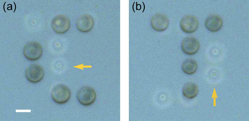
    Fig. 2. Optical image of double-layered silica spheres on a 30 nm thick Si3N4 membrane. The silica spheres were arranged in the form of (a) “C” and (b) “T” (b) on two sides of the membrane. The yellow arrows indicate spheres on the other side of the membrane. Scale bar, 10 μm.
    Experimental 2D diffraction patterns of the double-layered sample measured with the (a) green and (b) blue lasers. (c,d) The low spatial frequency regions of diffraction patterns (a,b) show that the missing intensity data are confined within the central speckles.
    Fig. 3. Experimental 2D diffraction patterns of the double-layered sample measured with the (a) green and (b) blue lasers. (c,d) The low spatial frequency regions of diffraction patterns (a,b) show that the missing intensity data are confined within the central speckles.
    Front view of the oversampled diffraction patterns on the Ewald spheres with the (a) blue and (b) green lasers. (c) The spherical diffraction patterns for two input wavelengths on a 3D Cartesian grid. (d) A schematic of the cross section of the diffraction patterns projected on the Ewald spheres for the green and blue lasers.
    Fig. 4. Front view of the oversampled diffraction patterns on the Ewald spheres with the (a) blue and (b) green lasers. (c) The spherical diffraction patterns for two input wavelengths on a 3D Cartesian grid. (d) A schematic of the cross section of the diffraction patterns projected on the Ewald spheres for the green and blue lasers.
    (a,b) 3D image (amplitude and phase) of the double-layered sample reconstructed from the diffraction patterns shown in Fig. 3. The central reconstructed slices of the spheres in (c) the front “C” and (d) back “T” layers. The blue dashed circles indicate the regions with a relatively low intensity due to the overlapped area of the two spheres perpendicular to beam direction.
    Fig. 5. (a,b) 3D image (amplitude and phase) of the double-layered sample reconstructed from the diffraction patterns shown in Fig. 3. The central reconstructed slices of the spheres in (c) the front “C” and (d) back “T” layers. The blue dashed circles indicate the regions with a relatively low intensity due to the overlapped area of the two spheres perpendicular to beam direction.
    Jian Zhang, Jiadong Fan, Jianhua Zhang, Qingjie Huang, Huaidong Jiang. 3D imaging by two-color Ewald spheres with optical lasers[J]. Chinese Optics Letters, 2016, 14(11): 111102
    Download Citation