• Acta Optica Sinica
  • Vol. 40, Issue 4, 0410002 (2020)
Xiaowen Lü, Feng Shao*, Yiming Xiong, and Weishan Yang
Author Affiliations
  • Faculty of Electrical Engineering and Computer Science, Ningbo University, Ningbo, Zhejiang 315211, China
  • show less
    DOI: 10.3788/AOS202040.0410002 Cite this Article Set citation alerts
    Xiaowen Lü, Feng Shao, Yiming Xiong, Weishan Yang. Retinal Vessel Segmentation Method Based on Two-Stream Networks[J]. Acta Optica Sinica, 2020, 40(4): 0410002 Copy Citation Text show less
    Overall framework of proposed method
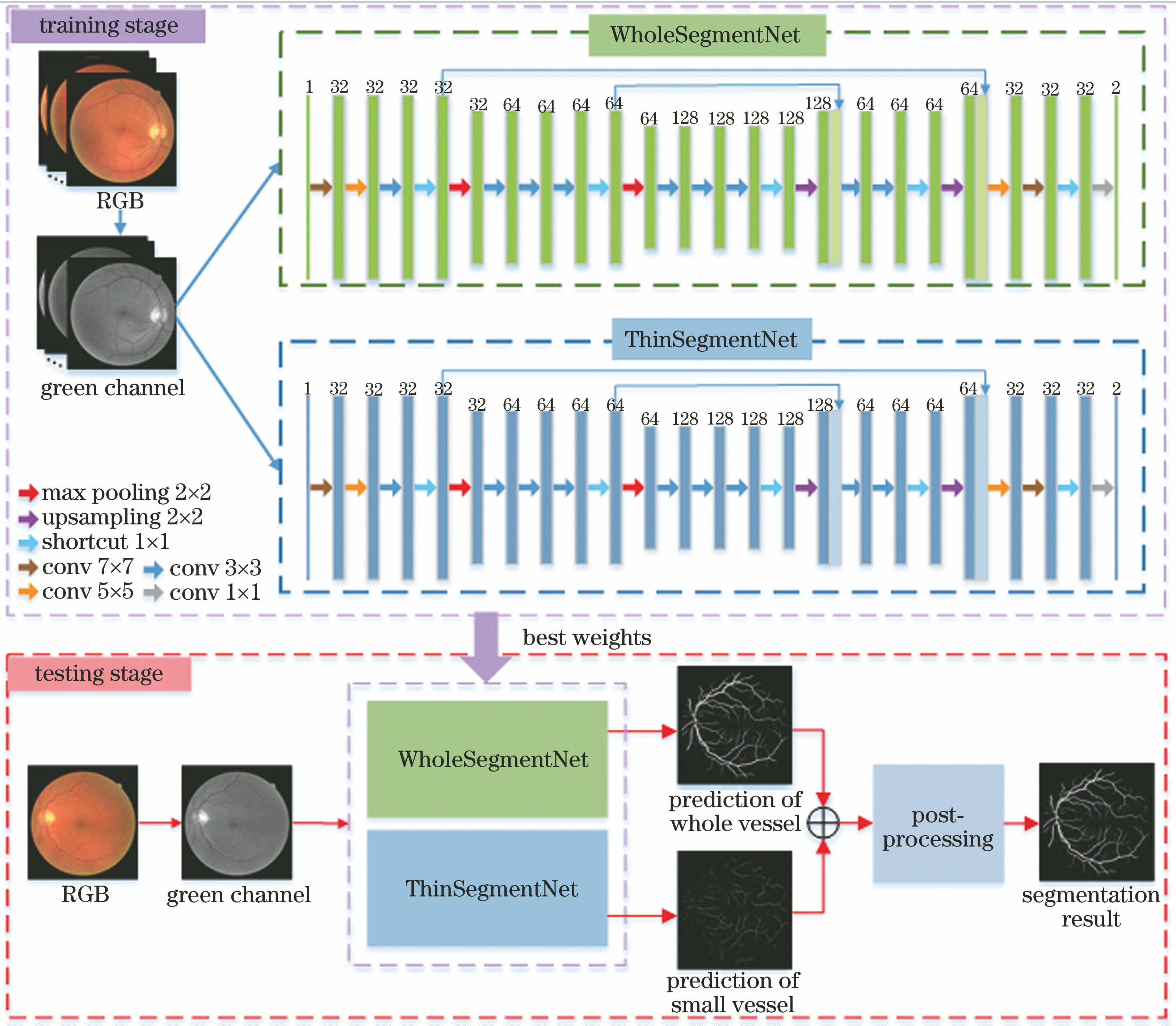
    Fig. 1. Overall framework of proposed method
    Input image and corresponding ground truth in training stage. (a) Input images; (b) ground truth for training WholeSegmentNet; (c) ground truth for training ThinSegmentNet (dark color area in picture)
    Fig. 2. Input image and corresponding ground truth in training stage. (a) Input images; (b) ground truth for training WholeSegmentNet; (c) ground truth for training ThinSegmentNet (dark color area in picture)
    Residual network structure
    Fig. 3. Residual network structure
    Results of database partitioning of DRIVE, STARE and CHASE_DB1. (a) Original images; (b) ground truth; (c) segmented whole vessel images; (d) segmented small vessel images; (e) fusion results; (f) results of proposed method
    Fig. 4. Results of database partitioning of DRIVE, STARE and CHASE_DB1. (a) Original images; (b) ground truth; (c) segmented whole vessel images; (d) segmented small vessel images; (e) fusion results; (f) results of proposed method
    ROC curves with different databases. (a) DRIVE; (b) STARE; (c) CHASE_DB1
    Fig. 5. ROC curves with different databases. (a) DRIVE; (b) STARE; (c) CHASE_DB1
    Effect of ThinSegmentNet segmentation and post-processing on segmentation results. (a) Ground truth; (b) vessel images of WholeSegmentNet predictions; (c) vessel images of WholeSegmentNet+ThinSegmentNet predictions; (d) results of proposed method
    Fig. 6. Effect of ThinSegmentNet segmentation and post-processing on segmentation results. (a) Ground truth; (b) vessel images of WholeSegmentNet predictions; (c) vessel images of WholeSegmentNet+ThinSegmentNet predictions; (d) results of proposed method
    Segmentation of vessels in different areas. (a) Segmentation of vessels in pathological areas; (b) segmentation of vessels in central line reflex areas; (c) segmentation of vessels in low contrast areas
    Fig. 7. Segmentation of vessels in different areas. (a) Segmentation of vessels in pathological areas; (b) segmentation of vessels in central line reflex areas; (c) segmentation of vessels in low contrast areas
    DatabaseMethodRseRspRAccAUC
    WholeSegmentNet0.75780.98160.95310.9732
    DRIVEWhole+ThinSegmentNet0.79710.97360.95220.9702
    Proposed method0.80620.97690.95470.9739
    WholeSegmentNet0.77190.98280.96100.9766
    STAREWhole+ThinSegmentNet0.80950.97570.95860.9737
    Proposed method0.83080.97840.95930.9788
    WholeSegmentNet0.76890.98010.96090.9778
    CHASE_DB1Whole+ThinSegmentNet0.79970.97310.96010.9764
    Proposed method0.81350.97620.96170.9782
    Table 1. Evaluation results of small vessel segmentation and post-processing methods in DRIVE, STARE and CHASE_DB1 test sets
    MethodDRIVESTARECHASE_DB1
    RseRspRAccAUCRseRspRAccAUCRseRspRAccAUC
    Ref. [22]0.73950.97820.94940.96720.73170.98420.9560.96730.76150.95750.94670.9623
    Ref. [5]0.76550.97040.94420.96140.77160.97010.94970.95630.75850.95870.93870.9487
    Ref. [23]0.74200.98200.95400.86200.78000.97800.95600.8740
    Ref. [8]0.77430.97250.94760.96360.77910.97580.95540.97480.76260.96610.94520.9606
    Ref. [9]0.75690.98160.95270.97380.77260.98440.96280.98790.75070.97930.95810.9716
    Ref. [10]0.78970.96840.7680.97380.72770.9712
    Ref. [24]0.76910.98010.95330.9744
    Ref. [14]0.76530.98180.95420.97520.75810.98460.96120.98010.76330.98090.96100.9781
    Proposed method0.80620.97690.95470.97390.83080.97840.95930.97880.81350.97620.96170.9782
    Table 2. Comparison of retinal vessel segmentation results among different methods on DRIVE, STARE and CHASE_DB1 databases
    MethodRseRspRAcc
    Ref. [18]0.65870.95650.9258
    Ref. [25]0.72620.97640.9511
    Ref. [9]0.78000.98050.9672
    Proposed method0.81370.97580.9609
    Table 3. Evaluation results of 10 pathological images in STARE database
    Xiaowen Lü, Feng Shao, Yiming Xiong, Weishan Yang. Retinal Vessel Segmentation Method Based on Two-Stream Networks[J]. Acta Optica Sinica, 2020, 40(4): 0410002
    Download Citation