Author Affiliations
1School of Medical Technology, Beijing Institute of Technology, Beijing 100081, China2Department of Laser Medicine, the First Medical Center, Chinese PLA General Hospital, Beijing 100853, Chinashow less
Fig. 1. Schematic of action principle of PDT
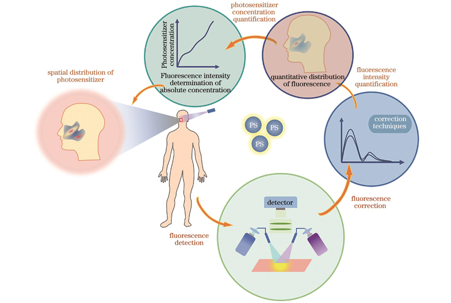
Fig. 2. Procedure for quantifying photosensitizer concentrations
Fig. 3. Factors affecting fluorescence intensity measurement of photosensitizers
Fig. 4. Instrumental factors affecting measurement of fluorescence intensity of photosensitizers
[23] Fig. 5. Intrinsic factors affecting measurement of fluorescence intensity of photosensitizers. (a) Tissue optical properties; (b) endogenous fluorescence
Fig. 6. Calibration algorithms for fluorescence spectra
Fig. 7. Schematics of quantitative detection techniques of photosensitizer. (a) Schematic of contact spectroscopic probe; (b) schematic of non-contact wide-field imaging system
Fig. 8. Schematics of contact spectroscopic probes for quantitative fluorescence detection. (a) Novel contact probe-based fluorescence dosimeter
[41]; (b)
in vivo photodynamic dosimeter probe using multi-excitation multi-emission system
[57]; (c)
in vivo fluorescence quantitative detection probe based on multi-spectrum
[43] Fig. 9. Schematics of non-contact wide-field imaging systems for quantitative detection of fluorescence intensity. (a) Quantitative spatial frequency domain fluorescence imaging
[32]; (b) snapshot quantitative fluorescence imaging system based on spatial frequency domain fluorescence imaging
[53]; (c) wide-field spectral-resolved fluorescence imaging system
[62]; (d) multimodal quantitative fluorescence imaging system
[3] | Technique | Classification | Ref. | Characteristic |
|---|
| Contact spectroscopic probe | - | [41][57] | 1) It’s more accurately to describe background signal and often used as a gold-standard.2) It is unaffected by topographic variations (as long as good contact with tissue surface is achieved)[70].3) It provides tissue imaging view fields at mesoscopic scales of order of one millimeter[3]. | | Non-contact wide-field imaging system | Quantitative spatial frequency domain fluorescence imaging | [67][68] | 1) Imaging allows for mapping spatial distribution of photosensitizer.2) Longer acquisition time of non-contact wide-field imaging compared with probe is susceptible to temporal changes occurring in surgical field during measurement[70].3) Fluorescence imaging is highly sensitive to lighting variations, due to irregular and non-flat tissue surfaces, and operating room environment[41]. | | Snapshot quantitative fluorescence imaging | [69][53] | | Wide-field spectrum differentiation fluorescence imaging | [62][70] | | Novel quantitative fluorescence imaging technique | Fluorescence tomography | [75][77] | Technology produces wide-field image estimates of depth of fluorophore[77]. | | Single-cell resolved microscopic fluorescence quantitative technique | [80][35] | Optical microscopy provides capabilities to view photosensitizer-fluorescing tumor regions at cellular resolution[80]. | | Portable quantitative fluorescence imaging technology | [29][84] | Quantitative wide-field fluorescence imaging system is low-cost and designed for maximal ease of translation into clinical workflow[84]. | | Endoscopic fluorescence quantitative imaging technology | [11][86] | Technology is least invasive and gives access to imaging of internal body organs[86]. |
|
Table 1. Quantitative detection technologies of fluorescence of photosensitizers
| Quantification technique | Quantification algorithm | Ref. | Photosensitizer | Precision | Time | Advantage | Limitation |
|---|
| QSFFI | Gardner
 s algorithm s algorithm | [32] | PpIX | 0.2 μg/mL | - | High resolution | To require many acquisition frequencies and extensive computation time | Kim
 s algorithm s algorithm | [67] | PpIX | 13 ng/mL | Slow | | 2-frequencyLUT | [68] | PpIX | ~5 μg/mL | ~1 min | | SQFI | Kim
 s algorithm s algorithm | [69] | ZW-800 | - | - | Fast imaging | Unable toseparate contributions of multiple fluorophores | Kim
 s algorithm s algorithm | [53] | ZW800-1 | - | Real-time | | WSRFI | Spectral constraint normalization | [70] | PpIX | 0.014 μg/mL | - | High detection sensitivity | To require extensive computation time | | Spectral constraint normalization | [71] | PpIX | 8 ng/mL | - | | Spectral constraint dual-band normalization | [40] | PpIX | 20 ng/mL | Near real-time | | Improved spectrally constrained normalization | [62] | PpIX | 10 ng/mL | Near real-time |
|
Table 2. Non-contact wide-field imaging technologies for quantitative detection of fluorescence intensity