• Chinese Journal of Lasers
  • Vol. 50, Issue 21, 2107201 (2023)
Jianfeng Yan1, Jiayuan Li1, Xing Zhang2, Jun Tan1, Jinyu Fu1, Caifeng Ou3, Chengyun Zhang1, Yunfeng Luo2, Zhifeng Chen1、*, and Pusheng Zhang2、**
Author Affiliations
  • 1School of Physics and Materials Science, Guangzhou University, Guangzhou 510006, Guangdong, China
  • 2Department of Breast Surgery, Zhujiang Hospital of Southern Medical University, Guangzhou 510280, Guangdong, China
  • 3Department of Breast Care Surgery, The First Affiliated Hospital of Guangdong Pharmaceutical University, Guangzhou 510080, Guangdong, China
  • show less
    DOI: 10.3788/CJL230982 Cite this Article Set citation alerts
    Jianfeng Yan, Jiayuan Li, Xing Zhang, Jun Tan, Jinyu Fu, Caifeng Ou, Chengyun Zhang, Yunfeng Luo, Zhifeng Chen, Pusheng Zhang. Specific Identification of Breast Tumors Based on Laser-Induced Autofluorescence Spectroscopy[J]. Chinese Journal of Lasers, 2023, 50(21): 2107201 Copy Citation Text show less
    Experimental system for laser-induced steady fluorescence spectroscopy
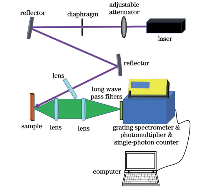
    Fig. 1. Experimental system for laser-induced steady fluorescence spectroscopy
    Laser-induced steady autofluorescence spectra of normal breast gland tissues and breast malignant tumor tissues. (a) Normal breast gland tissues; (b) breast malignant tumor tissues
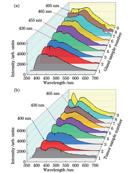
    Fig. 2. Laser-induced steady autofluorescence spectra of normal breast gland tissues and breast malignant tumor tissues. (a) Normal breast gland tissues; (b) breast malignant tumor tissues
    Identification effect of three fluorescence ratio methods on normal breast glands and breast malignant tumors. (a) I430/I465 fluorescence ratio method; (b) I430/I400 fluorescence ratio method; (c) 2I430/(I400+I465) fluorescence ratio method
    Fig. 3. Identification effect of three fluorescence ratio methods on normal breast glands and breast malignant tumors. (a) I430/I465 fluorescence ratio method; (b) I430/I400 fluorescence ratio method; (c) 2I430/(I400+I465) fluorescence ratio method
    Gaussian decomposed fittings of normal breast glands and breast malignant tumors spectra. (a) Normal breast glands spectra; (b) breast malignant tumors spectra
    Fig. 4. Gaussian decomposed fittings of normal breast glands and breast malignant tumors spectra. (a) Normal breast glands spectra; (b) breast malignant tumors spectra
    TissueFitting results
    Peak 1Peak 2Peak 3Peak 4
    FluorophoreCollagen+elastinNADHFlavin adenine
    Gland

    8.8%±1.2%

    389 nm±0.46 nm

    18.4%±3.3%

    416 nm±1.34 nm

    31.4%±8.4%

    465 nm±3.01 nm

    41.3%±7.9%

    532 nm±13.43 nm

    Tumor

    4.0%±0.6%

    384 nm±0.31 nm

    11.5%±0.9%

    401 nm±1.23 nm

    46.8%±6.8%

    459 nm±1.20 nm

    37.6%±8.0%

    537 nm±18.92 nm

    Table 1. Spectral fitting results and corresponding fluorophores
    Jianfeng Yan, Jiayuan Li, Xing Zhang, Jun Tan, Jinyu Fu, Caifeng Ou, Chengyun Zhang, Yunfeng Luo, Zhifeng Chen, Pusheng Zhang. Specific Identification of Breast Tumors Based on Laser-Induced Autofluorescence Spectroscopy[J]. Chinese Journal of Lasers, 2023, 50(21): 2107201
    Download Citation