Breast cancer ranks highest in global incidence among all cancers, posing a significant threat to women's health. During surgical treatment for breast cancer, the absence of swift and precise tissue identification often results in either removal of excessive tissue or extended surgical durations. Recently, autologous tissue spectroscopy has garnered interest as a detection method due to its benefits, including speed, sensitivity, selectivity, and non-invasiveness. Consequently, identifying effective spectral diagnostic features for breast cancer and understanding their mechanisms are critically important.
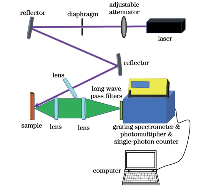
When normal tissue transforms into malignant tumor tissue, there is a qualitative shift in its internal biochemical environment. Hence, when normal glandular tissue undergoes carcinogenesis, the content and distribution of various fluorescent groups within that tissue alters, causing changes in the steady-state fluorescence spectral characteristics. In this study, a custom-built laser-induced steady fluorescence spectroscopy system was employed to conduct autofluorescence spectroscopy experiments on several malignant breast tumors and normal breast tissues (Fig. 1). A 355 nm sub-nanosecond sequence pulse laser served as the excitation light source. This choice was favorable for achieving a favorable signal-to-noise ratio at lower average optical power levels without inflicting irreversible damage to the samples. The constructed spectral system boasted a resolution as precise as 0.1 nm and spanned a spectral measurement range from 200 nm to 900 nm. The integration of a photomultiplier tube with a single-photon counter facilitated the measurement of faint spectral signals.
By comparing the spectral characteristics of the two types of tissue samples (Fig. 2), the spectral curves of tumors exhibit varying degrees of concave features near 430 nm, while all spectral curves of normal breast tissue samples show no concave features around 430 nm. This stands out as the most significant spectral characteristic difference between the two types of samples. We select 400, 430, and 465 nm as the spectral feature peaks and compare three ratio-based methods: I430/I465, I430/I400, and 2I430/(I400+I465). Based on this comparison, we propose that the fluorescence ratio method of 2I430/(I400+I465) serves as a feasible spectral-specific discrimination approach to differentiate between normal glandular tissue and breast tumors (Fig. 3). Using a discrimination threshold of 0.95, it is possible to realize 100% discrimination for all tested sample points.
Further spectral fitting analysis with Gaussian models shows that the experimental measurements of tumors and normal glandular tissue consist of four spectral components (Fig. 4). The autofluorescence of the breast tissue observed in the experiment predominantly stems from the contribution of four endogenous fluorescent substances: collagen, elastin, nicotinamide adenine dinucleotide hydrogen (NADH), and flavin adenine (Table 1). It is clear that the primary spectral characteristic difference in the autofluorescence spectra of the two types of tissues, as previously analyzed, namely, the spectral concavity observed near 430 nm in malignant breast tumors, primarily arises from the variation in peak 3. The heightened content of the reduced coenzyme NADH in malignant breast tumors when compared to normal breast tissue primarily explains this discrepancy. Moreover, a reduction in NADH-binding proteins leads to spectral variations in cancerous tissues.
In this study, a 355 nm sub-nanosecond sequential pulsed laser is utilized as the excitation source and autofluorescence spectral experiments are performed on both malignant breast tumor tissue and normal breast tissue with a custom-built laser-induced steady-state fluorescence spectroscopy system. By comparing the spectral characteristics of these tissue types, we propose a specific discrimination method based on the distinct concave features observed near 430 nm. By computing the fluorescence ratio of 2I430/(I400+I465), breast cancer tissues can be precisely identified. Thus, to recognize breast cancer tissue, it is sufficient to measure the fluorescence intensity at these three key wavelengths and compute the corresponding fluorescence ratio. Further analysis using Gaussian models indicates that the breast tissue's autofluorescence predominantly stems from four endogenous fluorescent substances. The spectral shifts in cancerous tissues mainly arise from an increase in the reduced coenzyme NADH and a decrease in NADH-binding proteins. The breast cancer spectral discrimination approach introduced in this study demonstrates notable specificity and is underpinned by a clear biological rationale, presenting a new benchmark for swift clinical detection, especially valuable for margin evaluation during breast-conserving surgeries.