• Acta Optica Sinica
  • Vol. 43, Issue 7, 0717002 (2023)
Guang Han1、2, Hao Feng1, Siqi Chen1, Zhe Zhao2、3, Jinhai Wang1、2, and Huiquan Wang1、2、*
Author Affiliations
  • 1School of Life Sciences, Tiangong University, Tianjin 300387, China
  • 2Tianjin Key Laboratory of Optoelectronic Detection Technology and System, Tianjin 300387, China
  • 3School of Electronics and Information Engineering, Tiangong University, Tianjin 300387, China
  • show less
    DOI: 10.3788/AOS221763 Cite this Article Set citation alerts
    Guang Han, Hao Feng, Siqi Chen, Zhe Zhao, Jinhai Wang, Huiquan Wang. Detection Method of Regional Cerebral Blood Flow Based on Interferometric Diffusing Speckle Contrast Imaging Technology[J]. Acta Optica Sinica, 2023, 43(7): 0717002 Copy Citation Text show less
    Schematic diagram of iDSCA technology
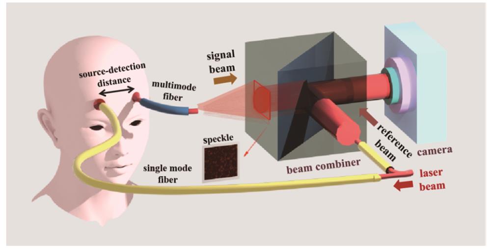
    Fig. 1. Schematic diagram of iDSCA technology
    Schematic diagram of hardware composition of iDSCA imaging system for detecting rCBF
    Fig. 2. Schematic diagram of hardware composition of iDSCA imaging system for detecting rCBF
    Hardware composition and model diagram of phantom velocity experiment. (a) Hardware composition of phantom velocity experiment; (b) phantom model of regional blood flow in cerebral cortex
    Fig. 3. Hardware composition and model diagram of phantom velocity experiment. (a) Hardware composition of phantom velocity experiment; (b) phantom model of regional blood flow in cerebral cortex
    Schematic diagram of in vivo experiment process. (a) Measurement mode of in vivo experiment; (b) picture of continuous noninvasive blood pressure monitor; (c) cuff-induced occlusion protocol
    Fig. 4. Schematic diagram of in vivo experiment process. (a) Measurement mode of in vivo experiment; (b) picture of continuous noninvasive blood pressure monitor; (c) cuff-induced occlusion protocol
    Schematic diagram of iDSCA speckle imaging results of phantom velocity experiment
    Fig. 5. Schematic diagram of iDSCA speckle imaging results of phantom velocity experiment
    Normalized fitting curve of multi-SD analysis varying with flow velocity. (a) Flow contrast; (b) coherence factor; (c) BFI
    Fig. 6. Normalized fitting curve of multi-SD analysis varying with flow velocity. (a) Flow contrast; (b) coherence factor; (c) BFI
    Normalized fitting curve of BFI varying with standard flow in multi-TD and multi-SD analysis. (a) SD of 6 mm; (b) SD of 8 mm; (c) SD of 10 mm; (d) SD of 12 mm
    Fig. 7. Normalized fitting curve of BFI varying with standard flow in multi-TD and multi-SD analysis. (a) SD of 6 mm; (b) SD of 8 mm; (c) SD of 10 mm; (d) SD of 12 mm
    Waveform comparison of BFI and BP signals in time domain and frequency domain. (a) Time domain; (b) frequency domain
    Fig. 8. Waveform comparison of BFI and BP signals in time domain and frequency domain. (a) Time domain; (b) frequency domain
    Schematic diagram of BFI signal waveforms in three states in cuff-induced occlusion protocol
    Fig. 9. Schematic diagram of BFI signal waveforms in three states in cuff-induced occlusion protocol
    SD /mmTD of 4.8 mmTD of 6.4 mmTD of 7.9 mm
    60.780.550.47
    80.830.580.47
    100.790.500.40
    120.790.580.47
    Mean BFI0.800.550.45
    Relative error /%2.044.985.80
    Table 1. Normalized fitting BFI results of different TD at multiple SD (flow is 100 mL·min-1)
    Guang Han, Hao Feng, Siqi Chen, Zhe Zhao, Jinhai Wang, Huiquan Wang. Detection Method of Regional Cerebral Blood Flow Based on Interferometric Diffusing Speckle Contrast Imaging Technology[J]. Acta Optica Sinica, 2023, 43(7): 0717002
    Download Citation