Author Affiliations
School of Electronic and Optical Engineering, Nanjing University of Science and Technology, Nanjing 210094, Jiangsu, Chinashow less
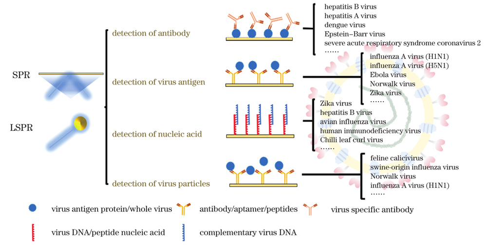
Fig. 1. Methods for virus detection based on SPR or LSPR technique
Fig. 2. Virus detection using AuNRs. (a) Detection principle of AuNRs nanosensor
[41]; (b) scanning electron microscopy (SEM) image of filter paper with AuNR-ZIKV-NS1 conjugate
[42]; (c) extinction spectra at different regions of BPD
[42]; (d) activity changes of ZIKV-NS1 on BPD under different storage conditions
[42] Fig. 3. Working diagrams of LSPR biosensor chip
[59]. (a) Schematic of virus detection by heteroassembled AuNPs chip; (b) schematic of sandwich structure composed of AuNPs and heteroassembled AuNPs layer for virus detection
Fig. 4. Schematic of multi-functional DNA 3-way connected HAuSN sensor
[62] Fig. 5. Nanostructure of EBOV sensor and fluorescence sensing method
[64]. (a) Structural diagram of 3D nano-antenna LSPR sensor chip; (b) decomposition diagram of sandwich structure
Fig. 6. Schematic of immune-fluorescence biosensor magnified by LSPR
[65] Fig. 7. Enzymatic or non-enzymatic amplification using DNA technique
Fig. 8. AuNPs modification of W(Mo)S
2 film rich in GB and its SEM diagram
[84] Fig. 9. LSPR combined with QDs to enhance sensitivity. (a) Mechanism diagams of DENV detection using AuNPs and hairpin ssDNA-CdSeTeS QDs
[85]; (b) schematics of four NP-Qdot-MB biosensor probes for ZIKV detection
[86] Fig. 10. Sensors with sandwich structure. (a) Plasma sensor with gold layer-virus-gold nanorod sandwich structure
[97]; (b) plasma sensor with gold cup array-virus-gold nanoparticle sandwich structure
[98] Fig. 11. AuNP-QD sensing probe. (a) Schematic of fluorescence intensity of adjacent QDs enhanced by LSPR effect of AuNPs
[106]; (b) mechanism of virus detection using CdSeTeS QDs/AuNPs rigid sensing probe
[107]; (c) mechanism of virus detection with adjustable length of intermediate junction peptide
[108]; (d) fluorescence quenching of QDs after virus captured by CdZnSeS/ZnSeS QD-peptide-AuNP sensor
[108] Fig. 12. Fluorescence spectral bar diagrams of CdSe QDs in various CdSe QD-peptide-AuNP nano-conjugates
[109]. AuNPs with six different sizes of (a) 80 nm, (b) 60 nm, (c) 45 nm, (d) 35 nm, (e) 25 nm, and (f) 15 nm combined with peptide with six different lengths to produce different fluorescence intensity changes; (g) effect of AuNPs with concentrations of 10
12, 10
9, and 10
6 mL
-1 on nano-conjugates with other constant parameters
Fig. 13. Sensor antifouling and reuse scheme. (a) Schematic of protein modified by polymer brush
[111]; (b) trapping of magnetic particles on reusable SPR chip through external magnetic field
[112]; (c) removal of magnetic particles by external magnetic field opposite to trapping direction
[112] | Method of detection | Sensor configuration | Technique | Capture element | Virus | Virus target | Sample solution | LOD | Time | Ref |
|---|
| Detect antibody | Au film | SPR | HBsAg | HBV | Anti-HBsAg Ab | HBS-EP buffer | 0.00098 mg/L | - | [12] | | Au film | SPR | gC protein | DPV | DPV Ab | HBS | - | - | [33] | | Au film | SPR | DENV2 Ag | DENV2 | IgM | Serum | 2.125 pmol/L | - | [35] | | Au film | SPR | SARS-CoV-2 S protein | SARS-CoV-2 | IgG | Serum | 1.04 ng·mm-2 | 10 min | [34] | | SARS-CoV-2 N protein | 1.34 ng·mm-2 | | Au film | SPR | VP1 | HAV | IgM | Serum | 0.218 nmol/L | - | [32] | | Au nanospike | LSPR | SARS-CoV-2 S protein | SARS-CoV-2 | Anti-SARS-CoV-2 S Ab | Serum | 0.08 ng/mL | 30 min | [36] | | AuNPs | LSPR | S protein | SARS-CoV-2 | Auti-S mAb | S.B. | 10 pg/mL | 15 min | [37] | | AuNRs | LSPR | DENV1-4 E protein | DENV | Anti-DENV mAb | Serum | 1 pg | - | [41] | | AuNRs | LSPR | ZIKV NS1 | ZIKV | Anti-ZIKV NS1 IgG | Serum | 200 ng/mL | - | [42] | | PBS | 1 ng/mL | | Au film | SPR | HBsAg | HBV | Anti-HBsAg Ab | Serum | <0.002 IU/mL | 10 min | [111] | | Au film | SPR | EBV Ag | EBV | Anti-EBV Ab | Serum | - | 30 min | [110] | | Au film | SPR | PHEMAT film | HBV | HBsAb | Serum | 208.2 mIU/mL | 30 min | [114] | | Gold capped nanoparticle array | LSPR | GBP | AIV | Auti-AI Ab | Carbonate buffer | 1 pg/mL | - | [115] | | Au film | SPR | DENV Ag | DENV | IgM | Serum | - | 10 min | [116] | | Au nanohole arrays | LSPR | PA | Anthrax toxin | 14B7 scFv Ab | - | 1 nmol/L | 800 s | [117] | | Au film | SPR | Ag-BSA conjugate | DENV | IgM Ab | PBS | - | 15 min | [118] | | Au film | SPR | Proteins | H5N1 | H5N1 Ab | PBS | 193.3 ng/mL | 30 min | [119] | | Au film | SPR | HA proteins | H1N1,H3N2,B | Ab | Serum | <0.5 μg/mL | 1-2 h | [120] | | Detect antigen | Au film | SPR | Anti-HIV-1 p24 mouse monoclonal antibodies | HIV | HIV-1 p24 protein | PBS | Physisorbed:(27±1)nmol/L | - | [48] | | Chemical:(4.1±0.5)nmol/L | | Au film- magnetic particles | SPR | Anti-NP Ab | H1N1 | H1N1 NP | PBS | 30 ng/mL | ~25 min | [112] | | Au film | SPR | EBOV mAb3 | EBOV | EBOV-rNP | PBS | 0.5 pg/mL | - | [49] | | Au film-graphene | SPR | HIV-1 p24 Ab | HIV | HIV-1 p24 Ag | PBS | 10 ng/mL | - | [51] | | Au film | SPR | IgM | DENV | DENV E protein | PBS | 0.0001 nmol/L | - | [52] | | Au film | SPR | DENV2 E lgM | DENV | DENV2 E protein | PBS | 0.08 pmol/L | 8 min | [53] | | Au film | SPR | DENV3 E lgM | DENV | DENV3 E protein | PBS | 0.08 pmol/L | 8 min | [54] | | Au film/electrode | SPR | Anti-H5N1 Ab | H5N1 | HA protein | - | 300 pmol/L | - | [55] | | AuNPs monolayer | LSPR | Auti-HBsAg | HBV | HBsAg | PBS | 10 pg/mL | 10-15 min | [59] | | AuNPs monolayer / | PBS | 100 fg/mL | | AuNPs | Serum | 10 pg/mL | | Glass-AuNPs | LSPR | NV binding peptides | NV | NV capsid proteins | MEM and FBS | 0.1 ng/mL | - | [60] | | Glass-AuNPs | LSPR | S1 aptamer | SARS-CoV-2 | S1 protein | Buffer | 0.26 nmol/L | - | [61] | | ITO-HAuNPs | LSPR | DNA 3WJ bioprobe | AIV H5N1 | HA protein | PBS | 1 pmol/L | 10 min | [62] | | Chicken serum | 1 pmol/L | | Au film | SPR | HA protein | Influenza A | HA protein | Deionized water | 0.72 μg/mL | 30 min | [63] | | 3D nanoantenna arrays | LSPR | IgG | EBOV | EBOV sGP | Plasma | 220 fg/mL | - | [64] | | AuNPs/CdSeTeS QDs | LSPR | Anti-NS1 Ab | ZIKV | ZIKV NS1 | Deionized water | 1.28 fg/mL | - | [65] | | Optic fiber/AuNPs | LSPR | A-H1 Ab | S-OIV | rHA protein | PBS | 13.9 pg/mL | - | [105] | | AuNRs | LSPR | HBsAb | HBV | HBsAg | Tris buffers | 0.01 IU/mL | - | [121] | | Au film | SPR | Anti-PB1-F2 Ab | H1N1 | PB1-F2 polypeptide | - | 10 nmol/L | 10 min | [122] | | Detect nucleic acid | Au film | SPR | - | HBV | HBV DNA templates | - | 2 fg/mL | 17 min | [73] | | Au film | SPR | Biotinylated DNA | HIV | HIV-1 pol 174bp | PBS | - | - | [74] | | Au film | SPR | Hairpin probes/DDT | HIV | HIV DNA | TE | 48 fmol/L | 60 min | [77] | | AuNPs | LSPR | ChiLCV 862BM R | ChiLCV | ChiLCV ssDNA | TE | 1.0 μg/mL | 10 min | [69] | | AuNPs | LSPR | ASOs | SARS-CoV-2 | SARS-CoV-2 RNA | | 0.18 ng/μL | 10 min | [70] | | AuNP-ssDNA-QD | LSPR | DENV1-4 hairpin ssDNA | DENV | Complementary ssDNA | DEPC | DENV1:24.6 fmol/L | - | [85] | | DENV2:11.4 fmol/L | | DENV3:39.8 fmol/L | | DENV4:39.7 fmol/L | | AgNP-Qdot646-MB | LSPR | MB | ZIKV | ZIKV RNA | - | 7.6 copy/mL | - | [86] | | AuNP-Qdot646-MB | 2.9 copy/mL | | Au/AgNP-Qdot646-MB | 2.4 copy/mL | | AuAgNP-Qdot646-MB | 1.7 copy/mL | | AuNI | LSPR | Thiol-cDNA | SARS-CoV-2 | RdRp-COVID | - | 0.22 pmol/L | - | [87] | | AuNI | LSPR | Thiol-cDNA | SARS-CoV-2 | SARS-CoV-2 NA | Nuclease-free water | (0.1±0.04)pmol/L | 30 min | [88] | | CFP | (0.275±0.051)fmol/L | | Au-W(Mo)S2 film-AuNPs | SPR | cDNA | SARS-CoV-2 | RNA | PBS | 0.1 fmol/L | - | [84] | | Au film | SPR | Capture probe(C1) | AIV | AI-DNA | Deionized water | 0.46 μmol/L | - | [68] | | Au film | SPR | BSMV oligonucleotide | BSMV | BSMV RNA | MgCl2+EDTA | 14.7 pg/μL | 50 min | [123] | | Au film | SPR | RNase H | H1N1 | MicroRNA miR-29a-3p | Mix | 1 nmol/L | 1 h | [124] | | Detect virus particles | Au film | SPR | Ab653,Ab956 | FCV | FCV particles | Oyster matrix | - | 15 min | [92] | | Ag-Au film | SPR | Auti-HA7 mAb | H7N9 | H7N9 particles | PBS | 144 copy/mL | 10 min | [94] | | Nasal mucosa | 402 copy/mL | | ITO-Au nanopattern | LSPR | mAb 2G12 | HIV | HIV-1 VLPs | DMEM | 200 fg/mL | - | [20] | | Au film | SPR | - | H1N1 | H1N1 particles | PBS | 0.2 fg·mm-2 | - | [93] | | Au film/AuNR | SPR | Anti-S protein Ab | SARS-CoV-2 | SARS-CoV-2 particles | - | - | - | [97] | | Au nanocup array/AuNPs | LSPR | Anti-S protein mAb | SARS-CoV-2 | Pseudovirus particles | PBS | - | 15 min | [98] | | Au film | SPR | MIP | Bacteriophage | MS2 | PBS | - | - | [99] | | POF-Au film | SPR | MIP | SARS-CoV-2 | SARS-CoV-2 particles | NaCl | - | 10 min | [101] | | Al-Al2O3/Qdot-streptavidin | SPR | Anti-VLP mAb12A11 | NV | NV VLPs | PBS | 0.01 ng/mL | - | [104] | | AuNPs/CdSeTeS QDs | LSPR | Anti-HA Ab/anti-NA Ab | H1N1 | H1N1 particles | Deionized water | 0.03 pg/mL | - | [106] | | Serum | 0.4 pg/mL | | CdSeTeS | LSPR | Anti-Nov Ab | NoV | NoVLPs | Deionized water | 12.1×10⁻¹⁵ g/mL | 1 min | [107] | | QD-AuNP probe | | QD-peptide-AuNP probe | LSPR | Anti-Ha Ab | H1N1 | H1N1 particles | Deionized water | 17.02 fg/mL | - | [108] | | QD-peptide-AuNP probe | LSPR | Anti-Nov Ab | NoV | NoVLPs | Serum | 124 fg/mL | - | [109] | | Anti-influenza Ab | H1N1 | H1N1 particles | 14.6 fg/mL | | Au film | SPR | mAb | OMSV | OMSV particles | TE | 1.36 ng/μL | 30 min | [91] | | Au film | SPR | Anti-MCMV Ab | MCMV | MCMV virus | PBS | 1×10⁻⁹ | 30 min | [90] | | Au film | SPR | PAb/ mAb | PVY | PVY particles | - | 0.31 μg/mL | - | [125] | | AuNPs | LSPR | Anti-T7 Ab | Bacteriophage | T7 | - | 18 pmol/L | - | [126] | | AuNPs | LSPR | IgG | DENV | DENV particles | PBS | - | 5 min | [127] |
|
Table 1. SPR or LSPR based biosensor for virus detection

![Virus detection using AuNRs. (a) Detection principle of AuNRs nanosensor[41]; (b) scanning electron microscopy (SEM) image of filter paper with AuNR-ZIKV-NS1 conjugate[42]; (c) extinction spectra at different regions of BPD[42]; (d) activity changes of ZIKV-NS1 on BPD under different storage conditions[42]](/richHtml/zgjg/2022/49/15/1507401/img_02.jpg)
