
Yu Zhang, Jie Lian, Mingyang Wei, Qingfen Jiang, Chenlin Wang, Yueming Wang, Zhen Xu. Research Progress of Imaging Ellipsometry[J]. Laser & Optoelectronics Progress, 2022, 59(10): 1000001
- Laser & Optoelectronics Progress
- Vol. 59, Issue 10, 1000001 (2022)
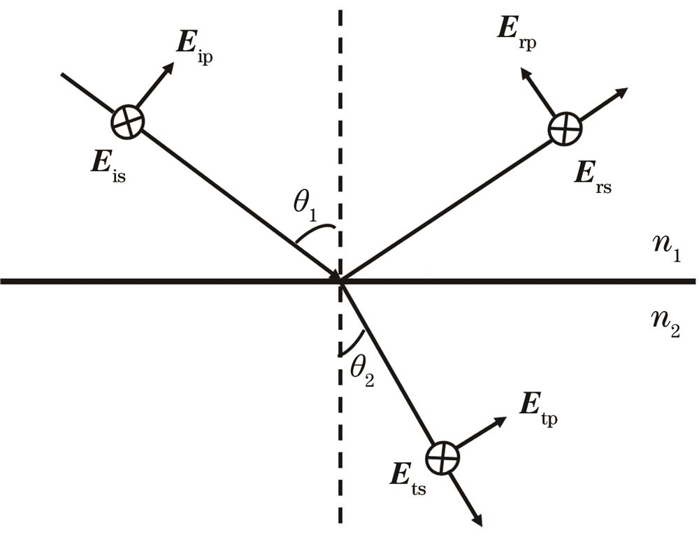
Fig. 1. Schematic of the reflection and refraction of monochromatic light on an isotropic and uniform material interface
![Schematic of magnification of imaging ellipsometer[14]](/richHtml/lop/2022/59/10/1000001/img_02.jpg)
Fig. 2. Schematic of magnification of imaging ellipsometer[14]
Fig. 3. Schematic of an imaging ellipsometer with an expanded beam, fixed polarizing components, and a CCD camera[8]
Fig. 4. Schematic of total reflection ellipsometer imaging system[36]
Fig. 5. Schematic of dual rotatingg compensator Mueller matrix imaging ellipsometer[4]
Fig. 6. Schematic of three-step phase shift imaging ellipsometer[48]
Fig. 7. Thickness profile of the SiO2 nanofilm on the Si substrate[48]
Fig. 8. Comparison of grayscale images of graphene sheets obtained by imaging ellipsometry and graphene sheets obtained by optical microscopy. (a) Optical micrograph of graphene sheets with different layers, the numbers in the figure represent the number of graphene layers; (b) ellipsometric grayscale image of graphene sheets on silica/silicon; (c)(d) ellipsometric Ψ diagram and Δ diagram of the box area in Fig.8(b) displayed at a higher resolution

Set citation alerts for the article
Please enter your email address
© Copyright 2018-2021 | Chinese Laser Press. All Rights Reserved 沪ICP备15018463号-20