• Advanced Photonics Nexus
  • Vol. 2, Issue 4, 046006 (2023)
Xuyang Chang1、2, Rifa Zhao1、2, Shaowei Jiang3, Cheng Shen4, Guoan Zheng5, Changhuei Yang4, and Liheng Bian1、2、6、*
Author Affiliations
  • 1Beijing Institute of Technology, MIIT Key Laboratory of Complex-Field Intelligent Sensing, Beijing, China
  • 2Beijing Institute of Technology, School of Information and Electronics and Advanced Research Institute of Multidisciplinary Sciences, Beijing, China
  • 3Hangzhou Dianzi University, School of Communication Engineering, Hangzhou, China
  • 4California Institute of Technology, Department of Electrical Engineering, Pasadena, California, United States
  • 5University of Connecticut, Department of Biomedical Engineering, Storrs, Connecticut, United States
  • 6Yangtze Delta Region Academy of Beijing Institute of Technology (Jiaxing), Jiaxing, China
  • show less
    DOI: 10.1117/1.APN.2.4.046006 Cite this Article Set citation alerts
    Xuyang Chang, Rifa Zhao, Shaowei Jiang, Cheng Shen, Guoan Zheng, Changhuei Yang, Liheng Bian. Complex-domain-enhancing neural network for large-scale coherent imaging[J]. Advanced Photonics Nexus, 2023, 2(4): 046006 Copy Citation Text show less
    Architecture of the proposed CI-CDNet. (a) Complex-domain neural network architecture. (b) Complex-domain convolution operation. (c) Multisource noise model for CI.
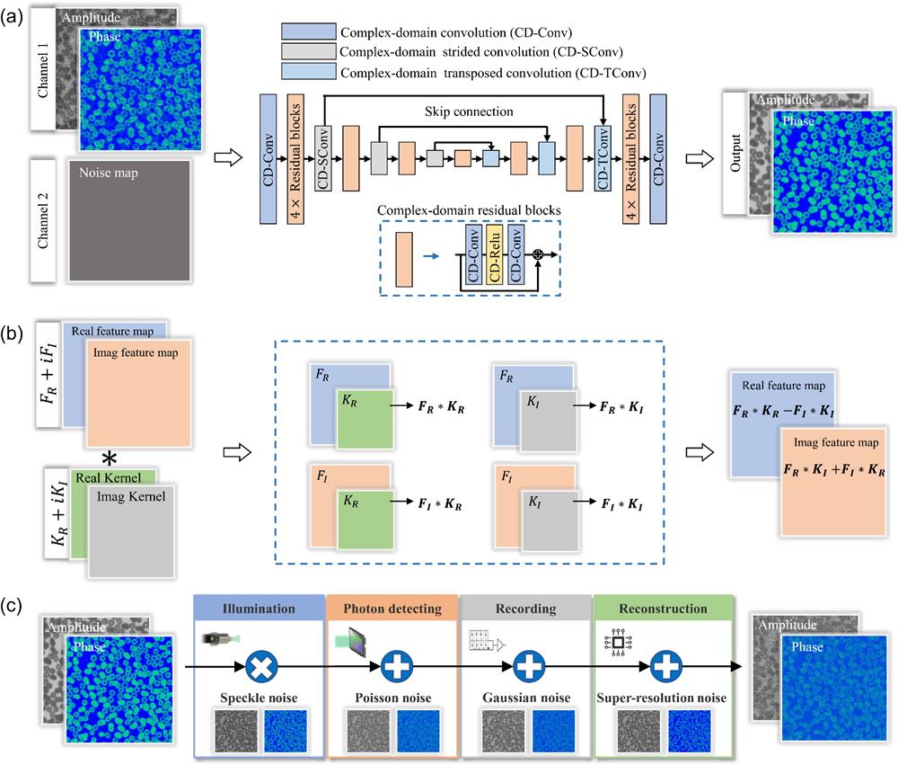
    Fig. 1. Architecture of the proposed CI-CDNet. (a) Complex-domain neural network architecture. (b) Complex-domain convolution operation. (c) Multisource noise model for CI.
    Experimental setup and resolution test of KKR holography under only 1 ms exposure time. (a) Experimental setup. CL, collimating lens; RL, relay lens; P, polarizer; BS, beam splitter; TL, tube lens; SLM, spatial light modulator. (b) Resolution test results of different enhancing methods using Siemens star under 1 ms exposure time. The blue and red curves are the cross sections of the images, which represent pixel-wise errors. The extremely short exposure time results in a low SNR of KKR direct reconstruction. (c) Running time (ms) of different enhancing methods.
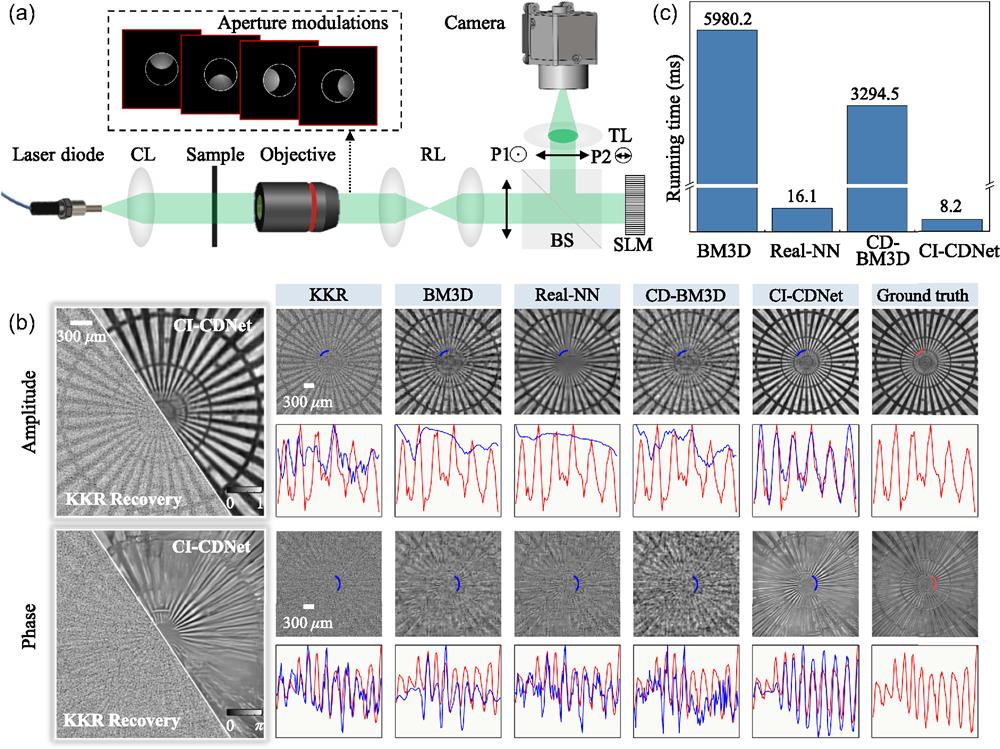
    Fig. 2. Experimental setup and resolution test of KKR holography under only 1 ms exposure time. (a) Experimental setup. CL, collimating lens; RL, relay lens; P, polarizer; BS, beam splitter; TL, tube lens; SLM, spatial light modulator. (b) Resolution test results of different enhancing methods using Siemens star under 1 ms exposure time. The blue and red curves are the cross sections of the images, which represent pixel-wise errors. The extremely short exposure time results in a low SNR of KKR direct reconstruction. (c) Running time (ms) of different enhancing methods.
    Quantitative results of KKR holography using a biological sample. (a) Enhancing results of different methods using a papillary thyroid carcinoma slide. (b) Quantitative results of CI-CDNet for reducing exposure time. The result of CI-CDNet under 1 ms exposure time is close to the results of KKR recovery under 50 ms exposure time (more results can be seen in Note 5 in the Supplemental Material).
    Fig. 3. Quantitative results of KKR holography using a biological sample. (a) Enhancing results of different methods using a papillary thyroid carcinoma slide. (b) Quantitative results of CI-CDNet for reducing exposure time. The result of CI-CDNet under 1 ms exposure time is close to the results of KKR recovery under 50 ms exposure time (more results can be seen in Note 5 in the Supplemental Material).
    Results of FPM under only 0.15 ms exposure time. (a) Experimental setup. (b) Resolution test of different enhancing methods using the USAF resolution test chart under 0.15 ms exposure time. (c) Enhancing results of the unstained blood smear under 0.15 ms exposure time. (d), (e) Quantitative results (blood smear) of amplitude and phase, respectively. The proposed CI-CDNet obtains more than 11 dB (amplitude) and 18 dB (phase) improvement on the PSNR index compared with the conventional AP method. (f) Running time (s) of different enhancing methods. AP is the baseline algorithm. Other enhancing methods are regularizers in FPM reconstruction; thus their running time includes the iteration time of data-fidelity term (Note 2 in the Supplemental Material).
    Fig. 4. Results of FPM under only 0.15 ms exposure time. (a) Experimental setup. (b) Resolution test of different enhancing methods using the USAF resolution test chart under 0.15 ms exposure time. (c) Enhancing results of the unstained blood smear under 0.15 ms exposure time. (d), (e) Quantitative results (blood smear) of amplitude and phase, respectively. The proposed CI-CDNet obtains more than 11 dB (amplitude) and 18 dB (phase) improvement on the PSNR index compared with the conventional AP method. (f) Running time (s) of different enhancing methods. AP is the baseline algorithm. Other enhancing methods are regularizers in FPM reconstruction; thus their running time includes the iteration time of data-fidelity term (Note 2 in the Supplemental Material).
    Results of LCP. (a) Schematic diagram of LCP system. (b), (c) Reconstructed amplitude and phase of the unstained blood smear using CI-CDNet with only 50 captured images. (d) Close-ups of three ROIs. The pseudo-color part is the phase and the gray part shows the amplitude. The GT is the result of ePIE using 900 images. (e) Results of white blood cell segmentation. (f) Results of virtual staining.
    Fig. 5. Results of LCP. (a) Schematic diagram of LCP system. (b), (c) Reconstructed amplitude and phase of the unstained blood smear using CI-CDNet with only 50 captured images. (d) Close-ups of three ROIs. The pseudo-color part is the phase and the gray part shows the amplitude. The GT is the result of ePIE using 900 images. (e) Results of white blood cell segmentation. (f) Results of virtual staining.
    AlgorithmData VolumeAmplitudePhase
    PSNRSSIMPSNRSSIM
    ePIE50 images16.380.4615.140.38
    100 images24.480.7321.670.50
    500 images30.010.9326.840.79
    CI-CDNet50 images30.590.8927.080.75
    Table 1. Quantitative results of LCP using different data volumes. The result of CI-CDNet using 50 captured images is close to the results of ePIE using 500 images (more results can be seen in Note 5 in the Supplemental Material).
    Xuyang Chang, Rifa Zhao, Shaowei Jiang, Cheng Shen, Guoan Zheng, Changhuei Yang, Liheng Bian. Complex-domain-enhancing neural network for large-scale coherent imaging[J]. Advanced Photonics Nexus, 2023, 2(4): 046006
    Download Citation