
Penglong Ren, Shangming Wei, Pu Zhang, Xue-Wen Chen, "Probing fluorescence quantum efficiency of single molecules in an organic matrix by monitoring lifetime change during sublimation," Chin. Opt. Lett. 20, 073602 (2022)
- Chinese Optics Letters
- Vol. 20, Issue 7, 073602 (2022)
Abstract
1. Introduction
Organic molecules are attractive to both physicists and chemists because molecules could have high quantum efficiencies in light emission and be chemically synthesized to have transitions at desired wavelengths. In the past several decades, single molecules embedded in solids, as isolated individual quantum systems, have become an attractive class of sources of single photons since a single two-level system cannot emit two photons simultaneously, as each excitation and emission cycle requires a finite time[
The quantum efficiency of an emitter indicates the ability to emit a photon once an excitation photon is absorbed and is defined as
In this work, we present a simple method to experimentally probe fluorescence quantum efficiency of single DBT molecules embedded in AC microcrystal by monitoring the fluorescence lifetime change during the process of natural sublimation. The decrease of the thickness of the microcrystal due to sublimation induces the change of the optical environment of the molecules and, consequently, the change of the Purcell factor or the local density of optical states (LDOS), which manifests through the modification of the fluorescence lifetime[
Sign up for Chinese Optics Letters TOC. Get the latest issue of Chinese Optics Letters delivered right to you!Sign up now
2. Experiments
DBT-doped AC (DBT:AC) microcrystals with preset concentrations are prepared through a co-sublimation process[
![]()
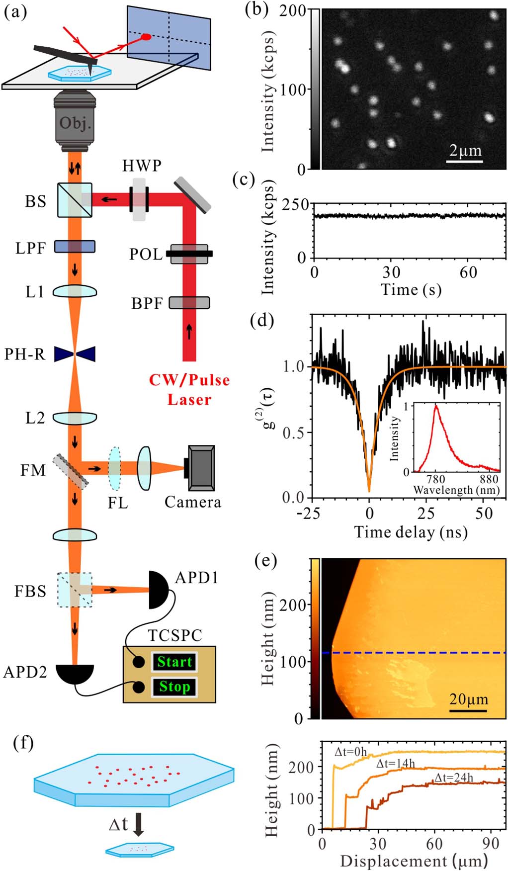
Figure 1.(a) Sketch of the experimental setup (see text for details). (b) Fluorescence image of single dibenzoterrylene (DBT) molecules embedded in an anthracene (AC) microcrystal obtained through confocal scanning; (c) photoluminescence (PL) time trace and (d) normalized second-order photon correlation function of fluorescence from a DBT molecule. The inset in (d) is the emission spectrum of the same molecule. (e) Atomic force microscope (AFM) topographic image of a part of an AC microcrystal. (f) Left: schematic illustration of the sublimation process of the AC microcrystal; right: cross-sectional plots of the height along the same blue dashed line shown in (e) at different times, which demonstrate the sublimation process.
3. Results
Figure 1(b) shows a confocal-scanning fluorescence image of single DBT molecules embedded in an AC microcrystal in a small region under CW laser excitation at a wavelength of 730 nm. Each molecule is spatially well separated and indicates a proper concentration of molecules in the sample. Figures 1(c) and 1(d) present a recorded PL time trace and normalized second-order photon correlation function
The structure of the sample we measured is depicted in Fig. 2(a), where
![]()
Figure 2.(a) Schematic diagram of the sample structure. (b) Measured and simulated back-focal plane (BFP) images of the emission from a single molecule oriented along the b axis of the AC microcrystal. (c)–(f) Measured and fitted PL decay curves from the same DBT molecule at different times. Insets: AFM topographic images of the region of the AC microcrystal where the molecule is located. The red star marks the location of the measured molecule. Scale bar: 500 nm.
Two different DBT molecules, molecule #1 and another labeled as molecule #2, are then studied in comparison. The crystal thickness-dependent total decay rates (
![]()
Figure 3.(a) Measured and fitted total decay rates (1/τ) of two different DBT molecules as functions of the AC microcrystal thicknesses. (b) Purcell factor distribution versus emitter dipole position h and the microcrystal thickness H.
Here,
During the sublimation, the position
Equation (3) provides a model to explain the sublimation-induced lifetime change. The unknown parameters
4. Discussion
We utilized the natural sublimation of the AC microcrystal, which induces optical environment change for embedded DBT molecules and experimentally probed fluorescence quantum efficiency of single DBT molecules by monitoring the fluorescence lifetime change due to the optical environment variation. By identifying the orientation of the molecule emission dipole from the radiation pattern through BFP imaging, we established a Purcell factor distribution as a function of crystal thickness and molecule position to describe the sublimation-induced lifetime change and analyze the quantum efficiency. Based on a series of measured AC crystal thicknesses and lifetimes recorded on the same molecules, we deduced the near-unity intrinsic quantum efficiency of the DBT molecule in the AC microcrystal.
References
[1] T. Basché, W. Moerner, M. Orrit, U. Wild. Single-Molecule Optical Detection, Imaging and Spectroscopy(1997).
[2] B. Lounis, M. Orrit. Single-photon sources. Rep. Prog. Phys., 68, 1129(2005).
[3] P. Kok, W. J. Munro, K. Nemoto, T. C. Ralph, J. P. Dowling, G. J. Milburn. Linear optical quantum computing with photonic qubits. Rev. Mod. Phys., 79, 135(2007).
[4] S. Scheel. Single-photon sources–an introduction. J. Mod. Opt., 56, 141(2009).
[5] S. V. Polyakov, A. L. Migdall. Quantum radiometry. J. Mod. Opt., 56, 1045(2009).
[6] P. Zhang, L. Lu, F. Qu, X. Jiang, X. Zheng, Y. Lu, S. Zhu, X.-S. Ma. High-quality quantum process tomography of time-bin qubit’s transmission over a metropolitan fiber network and its application. Chin. Opt. Lett., 18, 082701(2020).
[7] P. Michler, A. Kiraz, C. Becher, W. V. Schoenfeld, P. M. Petroff, L. Zhang, E. Hu, A. Imamoglu. A quantum dot single-photon turnstile device. Science, 290, 2282(2000).
[8] J. C. Loredo, N. A. Zakaria, N. Somaschi, C. Anton, L. de Santis, V. Giesz, T. Grange, M. A. Broome, O. Gazzano, G. Coppola, I. Sagnes, A. Lemaitre, A. Auffeves, P. Senellart, M. P. Almeida, A. G. White. Scalable performance in solid-state single-photon sources. Optica, 3, 433(2016).
[9] X. Ding, Y. He, Z. C. Duan, N. Gregersen, M. C. Chen, S. Unsleber, S. Maier, C. Schneider, M. Kamp, S. Höfling, C.-Y. Lu, J.-W. Pan. On-demand single photons with high extraction efficiency and near-unity indistinguishability from a resonantly driven quantum dot in a micropillar. Phys. Rev. Lett., 116, 020401(2016).
[10] I. Aharonovich, S. Castelletto, D. A. Simpson, C. H. Su, A. D. Greentree, S. Prawer. Diamond-based single-photon emitters. Rep. Prog. Phys., 74, 076501(2011).
[11] I. Aharonovich, S. Castelletto, D. A. Simpson, C. H. Su, A. D. Greentree, S. Prawer. Diamond-based single-photon emitters. Rep. Prog. Phys., 74, 076501(2011).
[12] B. Zhao, Y. Dong, S. Zhang, X. Chen, W. Zhu, F. Sun. Improving the NV generation efficiency by electron irradiation. Chin. Opt. Lett., 18, 080201(2020).
[13] J. Hong, C. Jin, J. Yuan, Z. Zhang. Atomic defects in two-dimensional materials: from single-atom spectroscopy to functionalities in opto-/electronics, nanomagnetism, and catalysis. Adv. Mater., 29, 1606434(2017).
[14] C. Toninelli, K. Early, J. Bremi, A. Renn, S. Götzinger, V. Sandoghdar. Near-infrared single-photons from aligned molecules in ultrathin crystalline films at room temperature. Opt. Express, 18, 6577(2010).
[15] C. Polisseni, K. D. Major, S. Boissier, S. Grandi, A. S. Clark, E. A. Hinds. Stable, single-photon emitter in a thin organic crystal for application to quantum-photonic devices. Opt. Express, 24, 5615(2016).
[16] S. Wei, P. Ren, Y. He, P. Zhang, X.-W. Chen. Single-molecule-doped crystalline nanosheets for delicate photophysics studies and directional single-photon-emitting devices. Phys. Rev. Appl, 13, 064023(2020).
[17] A. A. Nicolet, C. Hofmann, M. A. Kol’chenko, B. Kozankiewicz, M. Orrit. Single dibenzoterrylene molecules in an anthracene crystal: spectroscopy and photophysics. ChemPhysChem, 8, 1215(2007).
[18] D. Wang, H. Kelkar, D. Martin-Cano, T. Utikal, S. Götzinger, V. Sandoghdar. Coherent coupling of a single molecule to a scanning Fabry–Perot microcavity. Phys. Rev. X, 7, 021014(2017).
[19] S. Pazzagli, P. Lombardi, D. Martella, M. Colautti, B. Tiribilli, F. S. Cataliotti, C. Toninelli. Self-assembled nanocrystals of polycyclic aromatic hydrocarbons show photostable single-photon emission. ACS Nano, 12, 4295(2018).
[20] J. Hwang, E. A. Hinds. Dye molecules as single-photon sources and large optical nonlinearities on a chip. New J. Phys., 13, 085009(2011).
[21] S. Grandi, M. P. Nielsen, J. Cambiasso, S. Boissier, K. D. Major, C. Reardon, T. F. Krauss, R. F. Oulton, E. A. Hinds, A. S. Clark. Hybrid plasmonic waveguide coupling of photons from a single molecule. APL Photonics, 4, 086101(2019).
[22] P. Türschmann, N. Rotenberg, J. Renger, I. Harder, O. Lohse, T. Utikal, S. Götzinger, V. Sandoghdar. Chip-based all-optical control of single molecules coherently coupled to a nanoguide. Nano Lett., 17, 4941(2017).
[23] D. Rattenbacher, A. Shkarin, J. Renger, T. Utikal, S. Götzinger, V. Sandoghdar. Coherent coupling of single molecules to on-chip ring resonators. New J. Phys., 21, 062002(2019).
[24] X. Brokmann, L. Coolen, M. Dahan, J. P. Hermier. Measurement of the radiative and nonradiative decay rates of single CdSe nanocrystals through a controlled modification of their spontaneous emission. Phys. Rev. Lett., 93, 107403(2004).
[25] B. C. Buchler, T. Kalkbrenner, C. Hettich, V. Sandoghdar. Measuring the quantum efficiency of the optical emission of single radiating dipoles using a scanning mirror. Phys. Rev. Lett., 95, 063003(2005).
[26] S. Castelletto, I. Aharonovich, B. C. Gibson, B. C. Johnson, S. Prawer. Imaging and quantum-efficiency measurement of chromium emitters in diamond. Phys. Rev. Lett., 105, 217403(2010).
[27] R. J. Walters, J. Kalkman, A. Polman, H. A. Atwater, M. J. A. de Dood. Photoluminescence quantum efficiency of dense silicon nanocrystal ensembles in SiO2. Phys. Rev. B, 73, 132302(2006).
[28] W. Xu, X. Hou, Y. Meng, R. Meng, Z. Wang, H. Qin, X. Peng, X.-W. Chen. Deciphering charging status, absolute quantum efficiency, and absorption cross section of multicarrier states in single colloidal quantum dots. Nano Lett., 17, 7487(2017).
[29] E. M. Purcell, H. C. Torrey, R. V. Pound. Resonance absorption by nuclear magnetic moments in a solid. Phys. Rev., 69, 37(1946).
[30] W. L. Barnes. Fluorescence near interfaces: the role of photonic mode density. J. Mod. Opt., 45, 661(1998).
[31] G. Chen, J. Zhu, X. Li. Influence of a dielectric decoupling layer on the local electric field and molecular spectroscopy in plasmonic nanocavities: a numerical study. Chin. Opt. Lett., 19, 123001(2021).
[32] A. A. Nicolet, P. Bordat, C. Hofmann, M. A. Kol’chenko, B. Kozankiewicz, R. Brown, M. Orrit. Single dibenzoterrylene molecules in an anthracene crystal: main insertion sites. ChemPhysChem, 8, 1929(2007).
[33] L. Novotny, B. Hecht. Principles of Nano-Optics(2012).
[34] I. Nakada. The optical properties of anthracene single crystals. J. Phys. Soc. Jpn., 17, 113(1962).
[35] L. A. Nakhimovsky, I. Joussot-Dubien, M. Lamotte. Handbook of Low-Temperature Electronic Spectra of Polycyclic Aromatic Hydrocarbons(1989).


Set citation alerts for the article
Please enter your email address

© Copyright 2018-2021 | Chinese Laser Press. All Rights Reserved 沪ICP备15018463号-20