Transparent glassy composites incorporating lead-free anti-perovskite halide nanocrystals enable ultrastable high-resolution X-ray imaging. Credit: Le, Huang, et al. doi 10.1117/1.AP.5.4.046002.
In the realms of material inspection, medical diagnostics, astronomical discovery, and scientific research, the demand for high-resolution and ultrastable X-ray imaging methods has ignited a fervent pursuit of innovative X-ray-responsive materials. These sought-after materials must possess exceptional qualities such as high X-ray attenuation, efficient scintillation, rapid light decay, and robust durability. Among them, lead-halide-based perovskites have emerged as a compelling contender due to their remarkable luminescence efficiency, superior X-ray attenuation capabilities, and short fluorescence lifetimes. However, their application in the scintillation field is hindered by the toxicity of heavy metal lead (Pb), low photon yield caused by self-absorption effects, and poor X-ray irradiation stability.
Breaking barriers: Lead-free anti-perovskite nanocrystals
To overcome these challenges, researchers have sought solutions in lead-free zero-dimensional (0D) metal halides, such as copper-, silver-, zirconium-, and manganese-based halides. These intriguing alternatives have shown promise as effective scintillators for X-ray detection and imaging, boasting high photon yields, diverse composition and structure options, and a unique luminescence mechanism known as self-trapped excitons (STEs). However, a major hurdle lies in the fabrication of these metal halides as thin films or wafers, resulting in subpar imaging resolution due to light scattering caused by large particles and crystal boundaries. Additionally, lead-free 0D metal halides face challenges related to poor stability, particularly in hot and humid environments.
In an astounding breakthrough reported in Advanced Photonics, researchers from South China University of Technology developed a pioneering approach that revolutionizes X-ray imaging. They accomplished high-resolution and ultra-stable X-ray imaging even in demanding conditions of high temperature and humidity. The key: lead-free Cs3MnBr5 anti-perovskite nanocrystals embedded within a glass matrix.
Unlike traditional perovskite materials, anti-perovskites possess a distinctive structure represented as [MX4]XA3 [A = alkali metal; M = transition metal; and X = chlorine (Cl), bromine (Br), and iodine (I)]. This unique configuration features a luminescence center, the [MX4]2- tetrahedron, nestled within a three-dimensional (3D) XA6 octahedral anti-perovskite skeleton. This structure significantly reduces the interaction of the luminescence center, fostering enhanced spatial confinement effects and ultimately yielding high quantum efficiency and luminescence stability.
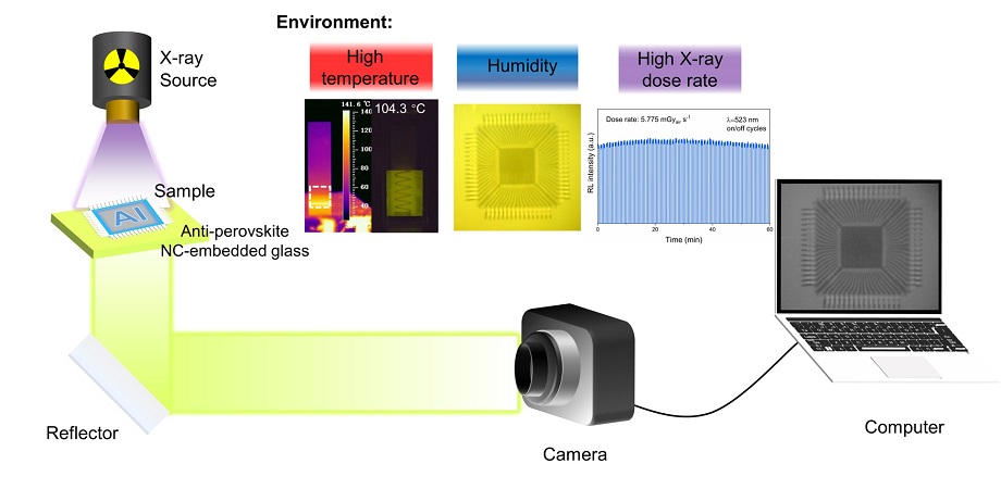
Through the process of in-situ crystallization during annealing, Mn2+ ions are seamlessly integrated into the glass matrix, giving rise to tunable luminescence colors ranging from red to green, as dictated by the annealing schedule. Moreover, the Cs3MnBr5 nanocrystal-embedded glass exhibits unparalleled X-ray irradiation stability, thermal stability, and water resistance. Remarkably, it also boasts an exceptional X-ray detection limit (767 nanograys per second), an impressive X-ray imaging spatial resolution (19.1 line pairs per millimeter), and outstanding X-ray dose irradiation stability (5.775 milligrays per second).

(a) Schematic of the X-ray imaging system. (b) Bright-field and X-ray images of the standard X-ray resolution pattern plate with the Cs3MnBr5 NC-embedded glass. (c) MTF of X-ray images obtained from the Cs3MnBr5 NC-embedded glass (the thickness is 0.6 mm). (d) Photographs of a cylindrical ABS resin embedded with an iron spring in air (top) and in dimethyl silicone oil (bottom). (e) Thermal imaging photographs (top) and X-ray images (bottom) of the cylindrical ABS resin embedded with an iron spring immersed in dimethyl silicone oil at different temperatures. Scale bar, 1 cm. (f) RL intensity of Cs3MnBr5 NCs in the glass recorded over continuous 120 on/off cycles during 60 min. (g) Photograph (left) and X-ray images (right) of the chip taken under continuous irradiation for 2 h. Scale bars, 2 mm. Credit: Le, Huang, et al. doi 10.1117/1.AP.5.4.046002.
This groundbreaking work presents an intriguing new scheme that harnesses the potential of transparent glassy composites incorporating lead-free anti-perovskite halide nanocrystals for high-resolution and ultrastable X-ray imaging applications. The results of this research could serve as a catalyst, stimulating further exploration and development of novel metal halide anti-perovskite materials. Ultimately, this discovery paves the way for the future development of next-generation X-ray imaging devices, promising transformative advancements in the field of X-ray diagnostics and imaging.
Read the Gold Open Access article by Le, Huang, et al., “Transparent glassy composites incorporating lead-free anti-perovskite halide nanocrystals enable tunable emission and ultrastable X-ray imaging,” Adv. Photon. 5(4) 046002 (2023), doi 10.1117/1.AP.5.4.046002