Photoacoustic imaging (PAI) uniquely combines the optical absorption contrast with ultrasound (US) detection. Owing to the much weaker US scattering by soft tissue than optical scattering, PAI has successfully broken the barrier in the imaging depth of high-resolution optical imaging. PAI not only has been widely used in life science and pre-clinical studies, but also is under rapid pace into clinical implementations. Among various PAI systems, the linear handheld probe-based system is one of the most commonly used, which is not only because of its simplicity and convenience, but also for its inherent capability to perform US imaging simultaneously. However, the short length of the traditional linear handheld probe strictly limits the view of the handheld probe. The limited view not only causes image artefacts, but also leads to failure in reconstruction of features due to the unavailability to detect Photoacoustic (PA) signals out of the view. This issue seriously affects the effective field of view of the PAI based on the linear handheld probe.
The research group led by Prof. Changhui Li from Peking University reported an easy method to effectively increase the total view angle of linear array aided by two acoustic reflectors in Chinese Optics Letters Vol. 19, No. 12, 2021(Guangjie Zhang, et al., Photoacoustic/ultrasound dual modality imaging aided by acoustic reflectors ).
The acoustic reflector setup is composed of two closely contacted right angle prisms, with their hypotenuse surfaces serve as two US reflectors. The acoustic reflectors equivalently turned the original single linear probe to three linear probes that can receive PA signals from three different directions. Part of the "V-shape" dip between the two reflectors is filled with a both acoustically and optically transparent agarose with a groove and the target is placed in the groove. To achieve acoustic coupling, ultrasound coupling agent will be filled into gap between imaging target and agarose platform during imaging, also an agarose mat will cover the target. The laser light can penetrate the transparent prism and agarose for stimulation, as shown in Figure 1.
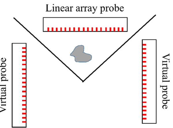
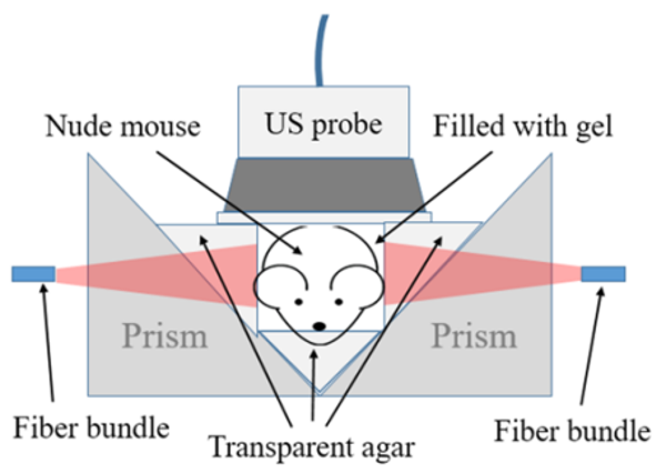
Fig. 1. Schematic mechanism of acoustic reflectors for PAI. (a) System setup; (b) In vivo imaging setup
Both phantom and in vivo animal study results demonstrated the acoustic reflector is a simple way to effectively retrieve those lost PA signals that cannot be detected by a traditional linear probe and thus substantially improve the PA image reconstruction. In addition, it is worth noting that the US reflector could not only help PAI, but also help US imaging itself, since the mirrored US images also. The proposed method has many clinical implementation potentials, including PAI of fingers, arms, and legs.