Surface Plasmon Resonance (SPR) is a prominent optical phenomenon that arises as the extent of energy transferring from photons to surface plasmon waves under appropriate conditions. In the past few years, this optical effect, owing to its high sensitivity, real-time detection, and anti-interference has already been extensively investigated and applied in medical treatment, environment monitoring, biomedical sensing and so on. Based on the principle of SPR, a novel D-shaped gold surface plasmon resonance photonic crystal fiber with one open-ring is proposed for detecting low refractive index materials has been investigated in detail. The proposed photonic crystal fiber of the simulation model is composed of three layers of air holes. The radii of air holes in the first-layer and third-layer are r1 and r3, respectively. While the second-layer air ring consists of air holes with two different radii, r2 and rs. The refractive index of air is fixed at nair = 1 and the radius of the cladding is R. A thin gold film with thickness tg is deposited on the inner surface of the micro-opening analyte channel on the upper side, the radius and the central location of the channel are rs and 2.5×Λ-1.25×rs, respectively. The fiber material is fused silica and the RI is determined by the Sellmeier equation, the relative dielectric constant of gold can be demonstrated by the Drude-Lorentz model. This paper uses the finite element method and sets the boundary conditions of the perfect matching layer for simulation. In order to investigate how the sensing performance of the proposed PCF-SPR sensor is affected by the parameters of the optical fiber, the effect of various parameters of the fiber such as air radii (r1, r2, r3, rs), air hole spacing (Λ) and the gold film (tg) on the SPR loss spectrum have been studied separately. The simulation results show that the confinement loss decreases as r1 increases. This can be attributed to the fact that more energy is confined to the core when r1increases, which affects the coupling between the core and plasmonic modes. At the same time, the confinement loss also decreases with the increase of r2, and the corresponding blue shift occurs with the resonance peaks moving toward a shorter wavelength over the process. The reason is that the increase of r2 will increase the refractive index difference between the plasmonic mode and core mode, which will affect the coupling between them. Therefore, with the increase of r2, the shorter wavelength can excite the plasmonic mode, resulting in the phenomenon of wavelength blue shift in the loss spectrum. Since the air holes of the third layer are located at the outermost part of the fiber, the change of r3 has little impact on the confinement loss, which can greatly reduce fabrication difficulty of the sensor. The pitches between the air holes are also an important factor in confinement loss, the change of Λ will influence the refractive index of core mode and plasmonic mode, which in turn affects the phase matching condition and energy coupling between them. The thickness of gold film plays a vital role in the sensing performance. If the gold film is too thick, the electric field can not penetrate the gold film, which will reduce the sensitivity of the proposed sensor. While if the gold film is too thin, the plasmonic wave will be strongly suppressed due to radiation damping. Therefore, the thickness of gold film can significantly affect the coupling between the core mode and the plasmonic mode. After optimizing the various parameters affecting the sensing performance of the sensor, we analyse the analytes with different refractive indices. Simulation results show that the sensor operates in the near-infrared and mid-infrared region with the wavelength range of 2 020~3 036 nm in the refractive index range of the analyte of 1.18~1.30. When the refractive index of the analyte is in the range of 1.23 to 1.30, the sensor operates in the band of 2 135~3 036 nm, and the average value of spectral sensitivity is up to 11 650 nm/RIU. When the refractive index of the analyte is between 1.29 and 1.30, the sensor operates in the mid-infrared band of 2 648~3 036 nm, and the maximum spectral sensitivity and resolution are 38 800 nm/RIU and 2.37×10⁻⁶ RIU, respectively. The proposed sensor shows great significance in detecting low refractive indexes in near- and mid-infrared waveband, and has potential applications in biomedical sensing, water environment and humidity detection and so on.
Surface Plasmon Resonance (SPR)is a prominent optical phenomenon that arises as to the extent of energy transferring from photons to surface plasmon waves under appropriate conditions[1]. In the past few years,this optical effect,owing to its high sensitivity,real-time detection,and anti-interference,has already been extensively investigated and applied in the fields of medical treatment[2],environment monitoring[3],biomedical sensing[4] and so on. The earliest SPR sensing devices were based on prism[5],such as the Kretschmann-Raether prism[6],whose sensitivity reached up to 10-6 RIU. Even though the prism can achieve high sensitivity,it has the disadvantage of costly integration,limited mechanical reliability and difficulties in mass production,which were limited to use in remote monitoring. Optical fiber-based SPR sensors have been exploited in recent years due to their advantages of easy miniaturization,low manufacturing cost and in-situ monitoring[7],etc. Especially,the sensors based on Photonic Crystal Fiber (PCF)can be easy phase-matching and encapsulation because of the minimized size,tunable geometric parameters and flexible design[8],so it becomes a promising candidate for assembling compact and ultra-sensitive SPR sensors with fast,sustainable and real-time monitoring.
As is well known that the sensing performance of Photonic Crystal Fiber-surface Plasmon Resonance (PCF-SPR)sensors depends on the metallic materials and the structure of PCF. Among the various plasmonic materials,gold[9] and silver[10] are mostly used due to their relatively low loss in the visible and near-infrared region,which makes it more effective to monitor the high energy transfer on the surface of plasmonic materials. To date,much effort has been devoted to the structure design of PCF to enhance the sensing performance of PCF-SPR sensors[11],and considerable variety of PCF-SPR structures have been reported,which can be divided into two categories,internal metal-coated structures using selectively coating or nanowires filled,and external metal-coated structures such as D-shaped,slotted and exposed core PCFs[12]. Among them,D-shaped PCF has attracted many interests compared with other PCF structures,because it can not only provide a good solution to the difficulties in a uniform coating which means the precise control of the thickness and roughness of the metal film on the outer surface of the optical fiber,but also solve the problem of filling analytes to Nano-size holes[13]. For example,AN Guowen,et al[14] proposed a D-shaped PCF-SPR sensor with a triangular lattice and four large-size channels,which can obtain the maximum wavelength sensitivity of 10 493 nm/RIU at 1.38. SINGH S and PRAJAPATI Y[15]presented a D-shape PCF-SPR sensor with gold-graphene layers on the surface. The sensitivity is 33 500 nm/RIU and the effective RI resolution is as high as 2.98×10⁻⁵ RIU for analyte RIs between 1.32 and 1.40. It can be seen from these reported literatures that the most reported D-shaped SPR sensors exhibit good sensing performance when the Refractive Index (RI)of the analyte is higher than 1.30.
However,with the development of biomedicine and material chemistry,the detection for low refractive index analyte has been paid more attention. For example,the RI of sevoflurane in the drug is around 1.27[16],the refractive index of some fluorine-containing organics compounds in water pollution is about 1.30[17]. Hence,it is essential for us to exploit sensors with high sensitivity for detecting low RI. In recent years,some research work about the RI of analytes lower than 1.30 has been conducted. For example,WANG Famei et al[18] studied a D-shaped sensor with two parallel to detect low RI ranging from 1.27 to 1.32 with the maximum spectral sensitivity up to 13 500 nm/RIU and the resonance wavelength ranging between 720 nm and 1 680 nm. LIU Chao et al[19] reported a mid-infrared PCF-SPR sensor with RI detection range of 1.23~1.29 and average sensitivity of 5 500 nm/RIU. CHEN Xin et al[20] proposed a D-type PCF-SPR sensor which operates in the mid-infrared band from ranging 2 530 nm to 2 750 nm with RI of 1.26~1.29 and the maximum sensitivity of 11 055 nm/RIU. In most of these studies,the reports of PCF-SPR sensor with operating wavelength in mid-infrared range are relatively few. In fact,SPR sensors operating in the mid-infrared wavelength have unique advantages compared to the visible and near-infrared bands[21]. For example,it is conducive to improve the detection accuracy and sensitivity of the sensor to large samples as living cells[22],and avoid the light damage to the biological samples[23]. Therefore,it is necessary to exploit the PCF-SPR sensor with higher sensitivity in mid-infrared waveband.
Herein,a novel D-shaped PCF-SPR sensor for detecting low RI analytes is proposed and analyzed. The open-ring channel coated with gold film can not only reduce the coating area,but also simplify the fabrication process. Numerical results show that a maximum spectral sensitivity of 38 800 nm/RIU can be obtained when analyte RI is 1.30 with a high resolution of 2.37×10⁻⁶ RIU. The operating wavelength of this fiber sensor includes the near- and mid-infrared regions ranging from 2 020 nm to 3 036 nm. This proposed sensor is of significance in environmental engineering,biosensors and healthcare.
1 Design and analysis of the model
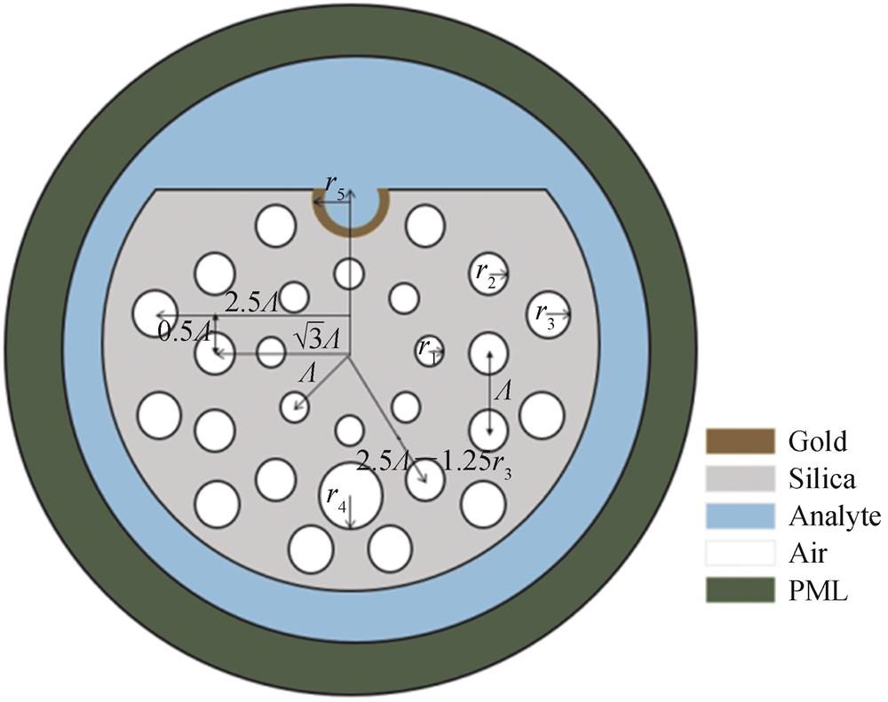
The cross-section of the proposed D-shaped SPR sensor is illustrated in Fig. 1. This structure contains three layers of air rings. The radii of air holes in the first-layer and third-layer are r1 and r3,respectively. While the second-layer air ring consists of air holes with two different radii,r2 and rs. The refractive index of air is fixed at nair = 1 and the radius of the cladding is R. A thin gold film with thickness tg is deposited on the inner surface of the micro-opening analyte channel on the upper side,the radius and the center location of the channel are rs and 2.5×Λ-1.25×rs,respectively. The first layer of air holes is composed of regular octagonal air holes with a radius of r1 and a distance of Λ from the fiber core,the second layer of air holes consists of a large air hole with radius of rs and ten small air holes with radius of r2. The large air hole is located directly below the fiber core and the distance from the fiber core is 2.5×Λ-1.25×rs,while the small air holes with radius of r2 are arranged in two ways. One is distributed on both sides of the large air hole and the distance from the fiber core is 2.5×Λ-1.25×rs,it is composed of four air holes with a radius of r2 which are 30 degrees from the vertical center line. The remaining six air holes are left-right symmetrical structure,in which the abscissa of the three air holes on the right is ,the distance between the three adjacent air holes on the right is Λ,and the remaining three air holes on the left are symmetrically distributed with the three air holes on the right. The outermost air holes are obtained by rotating the air holes with a radius of r3 with an abscissa of 2.5Λ and an ordinate of 0.5Λ for three times,and each rotation angle is 30°. Then four air holes will be obtained with a radius of r3,while the four air holes in the outermost layer on the left are symmetrically distributed with the four air holes with a radius of r3 on the right. The air hole structure of the proposed sensor is symmetrical so as to enlarge the energy leakage from the fiber core to enhance the coupling between optical and plasmonic modes.
Figure 1.Schematic illustration of the proposed D-shaped PCF-sensor
The D-shaped sensors can be fabricated by stack-and-draw method[24] and side-polishing technique[25],the gold film can be coated on the inner surface of the micro-opening channel by chemical vapor method[26]. The fiber material is fused silica and the RI is determined by the Sellmeier equation[27]
where represents the operating wavelength. Furthermore,the relative dielectric constant of gold can be demonstrated by Drude-Lorentz model,which is expressed as follows[28]
where is the permittivity of gold at high frequencies, is the plasma frequency of gold,and is the scattering frequency of electrons,and is the angular frequency of the incident light.
In order to prevent the light reflects from the PCF boundary,an artificial boundary condition known as the Perfectly Matched Layer (PML)[29] is added to absorb the radiation energy at the outermost layer. SPR sensor is based on the coupling between the core mode and the plasmonic mode,a loss peak occurs at resonance wavelength. The coupling strength between the fiber core mode and the plasmonic mode is described by the confinement loss of the core mode,which is expressed as[30]
π
where represents the confinement loss, is the imaginary part of the complex effective refractive index.
2 Results and discussion
Fig. 2 shows the relationship between the real part of the effective refractive index of core mode and plasmonic mode and the variation of confinement loss at a wavelength ranging from 1 800 nm to 2 300 nm at RI=1.2,in which the green curve stands for core mode,while the black curve represents the plasmonic mode. Meanwhile,the relationship between the confinement loss of the core mode and wavelength is represented by the red curve. As shown in Fig. 2,when the real part of the effective refractive index of the core mode and the plasmonic mode are equal,a strong SPR effect can be observed with a prominent peak in the loss spectrum. In other words,a maximum energy transfers from the core mode to the plasmonic mode at the resonance wavelength.
Figure 2.Real part of effective refractive index (left axis)and confinement loss (right axis)of core mode and plasmonic mode
Fig. 3 shows the energy transfer process between core and plasmonic mode when the wavelength changes from 1 800 nm to 2 300 nm. As it can be seen from the figure,when the wavelength is away from the optimal phase matching point,most of the energy is concentrated in the core,while only a small portion of the energy leaks into the sensing interface. Obviously,the maximum energy transfers at the resonant wavelength,which is caused by the phase matching condition of core mode and plasmonic mode.
Structure
RI range
Max sensitivity /(nm·RIU-1)
Max resolution
Year
Ref.
D⁃shaped
1.23~1.29
5 500
7.69×10⁻⁶
2017
[19]
Dual⁃shaped
1.27~1.32
13 500
7.41×10⁻⁶
2018
[18]
D⁃shaped
1.20~1.29
11 055
9.05×10⁻⁶
2019
[20]
D⁃shaped
1.19~1.29
10 700
____
2019
[17]
Microchannel
1.20~1.33
12 000
8.3×10⁻⁶
2020
[16]
D⁃shaped
1.18~1.30
38 800
2.37×10⁻⁶
2022
This work
Table 1. Comparison of the proposed sensor with previous designs
In order to optimize the sensing performance,the air holes radii,the pitch and gold film thickness were investigated separately. The refractive index of the analyte is fixed at 1.2 and the structural parameters of the PCF are calculated by keeping the rest of the structural parameters constant. The initial structural parameters r1, r2, r3, rs of the PCF are 0.5 µm,0.6 µm,0.7 µm and 1 µm,respectively. The innermost air hole pitch is 2.4 µm and the thickness of the gold film is 50 nm. It is worth noting that the variation of air hole diameter affects the coupling intensity and phase matching condition of core mode and plasmonic mode,thereby also affecting the energy conversion between them.
It can be seen from Fig. 4(a)that the resonance peak gradually decreases from 229.43 dB/cm to 203.49 dB/cm as r1 changes from 0.5 µm to 0.54 µm,and the resonance wavelength shifts from 2 070 nm to a longer wavelength of 2 150 nm. This can be attributed to the fact that more energy is confined to the core when r1 changes from 0.5 µm to 0.54 µm,which affects the coupling between the core mode and the plasmonic mode. Fig. 4(b)describes the confinement loss decrease when radius r2 changes from 0.6 µm to 0.8 µm,and the corresponding blue shift occurs with the resonance peaks moving toward a shorter wavelength from 2 100 nm to 1 650 nm over the process. The reason is that the increase of r2 will increase the refractive index difference between the plasmonic mode and core mode,which will affect the coupling between them. Therefore,with the increase of r2,the shorter wavelength can excite the plasmonic mode,resulting in the phenomenon of wavelength blue shift in the loss spectrum.
Figure 4.The confinement loss affected by changing the radii of the air holes and the open-ring
Fig. 4(c)describes the relationship between the confinement loss and the radius r3. As can be seen from the figure,the variation of r3 has almost no effect on the resonance wavelength and the confinement loss,which means the air hole r3 has a slight impact on the coupling between the core mode and the plasmonic mode. So the precision requirement of the radius r3 is low and therefore the fabrication difficulty of the sensor can be greatly reduced. Fig. 4(d)shows the effect of the different open-ring channel radii rs on the confinement loss of the core mode. It can be seen that the confinement loss reduces from 229.43 dB/cm to 54.21 dB/cm as rs increases slightly from 1 µm to 1.2 µm and the resonance wavelength decreases from 2 070 nm to 1 550 nm. Therefore,the performance of the proposed sensor can be optimized by adjusting the size of the open-ring channel. In this case,we set r1=0.5 µm,r2=0.6 µm,r2=0.7 µm,and rs=1 µm,respectively,for the proposed sensor.
Fig. 5(a)depicts the variation of the confinement loss of the proposed sensor with different pitches ranging from 2.4 µm to 2.56 µm. It can be seen that when Λ increases,the resonance wavelength gradually decreasing from 2 070 nm to 1 870 nm with the resonance peak decreases from 229.40 dB/cm to 96.76 dB/cm. The reason is that the change of Λ has an impact on the refractive index of core mode and plasmonic mode,which in turn affects the phase matching condition and energy coupling between them. Meanwhile,if the pitch is too large,it will prevent the concentration of the energy in the core. Totally,the pitch of 3 µm is selected to prevent the gap from increasing.
Figure 5.The confinement loss of the proposed sensor varies with Λ and tg
The thickness of gold film plays a vital role in the sensing performance. If the gold film is too thick,the electric field can not penetrate the gold film,which will reduce the sensitivity of the proposed sensor. While if the gold film is too thin,the plasmonic wave will be strongly suppressed due to radiation damping. Therefore,the thickness of gold film can significantly affect the coupling between the core mode and the plasmonic mode. Fig. 5(b)exhibits the loss spectra variation as the thickness of gold film ranges from 40 nm to 60 nm. As shown in Fig. 5(b),the reduction of the confinement loss and the blue shift appears as the thickness of the gold film increasing. A similar phenomenon was also noted in the paper[31]. Therefore,the sensitivity of the sensor can be improved by optimizing the thickness of the gold film.
The amplitude sensitivity is a critical parameter for evaluating the quality of the proposed sensor,and it is defined by the following equation[32]
where represents the difference of the confinement loss of the adjacent analyte RI, denotes the overall confinement loss,and represents difference of the analyteRI.
The resonance wavelength is used to detect the refractive index of the analyte,which means the analyte has a significant effect on the coupling intensity between the core mode and the plasmonic mode. The wavelength dependence of the loss spectra and the amplitude sensitivity of the sensor for analytesRI ranging from 1.18 to 1.30 has been shown in Fig. 6(a)and Fig. 6(b),respectively. The resonance wavelength shifts to a longer wavelength and the peak of the confinement loss also increases gradually when the analyteRI increases from 1.18 to 1.30. This can be attributed to the incomplete coupling between the core mode and the plasmonic mode. As shown in Fig. 6(a),with the RI increasing,the confinement loss increases from 209.98 dB/cm to 579.23 dB/cm. We can notice that the maximum amplitude sensitivity is 68.13 RIU-1 from Fig. 6(b).
Figure 6.The increase of the confinement loss and amplitude sensitivity varies with wavelength for analyteRI from 1.18 to 1.3
Fig. 7 illustrates the fitted curve of the resonance wavelength with the analyte RI changing from 1.18 to 1.30. The inserted table shows the results of the polynomial fitting,the slope of the fitted curve represents the sensitivity of the proposed sensor. The adjusted R-square value of the fitting curve is 0.995 08,which means a high agreement of fitting. It also can be seen that the resonant point moves to longer wavelengths as the analyteRI increases,with a maximum spectral sensitivity of 388 00 nm/RIU at the analyte RI of 1.30.
Figure 7.The resonance wavelength varies with analyte RI from 1.18 to 1.30
The spectral sensitivity has equal significance to the amplitude sensitivity at estimating the performance of the PCF sensor. The spectral sensitivity is defined by the following equation[33]
where represents the difference of the adjacent resonance wavelength.
In addition to the spectral and amplitude sensitivities,the resolution is a critical parameter of the proposed sensor. The resolution is expressed as[34]
where is the minimum spectral resolution. A high resolution of 2.37×10⁻⁶ RIU can be gained at 1.30 in the proposed structure as shown in Table 1.
Table 1 shows the performance of the proposed sensor compared to previous sensors. It can be seen that the sensor proposed in this work has the advantage of higher refractive index detection. Meanwhile,the detection range of the proposed sensor is wider than that of the previous sensor.
3 Conclusion
In this work,a novel D-shaped PCF sensor based on SPR effect is proposed and numerically investigated. Unlike conventional D-shaped structures,the SPR effect is excited by coating in the open-ring channel with a thin gold film,which can enhance the resonance effect and improve the spectral sensitivity of proposed sensor. In addition,the usage of this channel reduces the coating area of gold film. This makes the fabrication process more simplified and cost-effective. Simulation results show that the sensor can detect low refractive indexes ranging from 1.18 to 1.30 with the highest spectral sensitivity and resolution of 38 800 nm/RIU and 2.37×10⁻⁶ RIU. Due to the low refractive index sensing range and the simple compact fiber design,the proposed SPR sensor could be a competitive candidate in low refractive index detection.