• Chinese Journal of Lasers
  • Vol. 47, Issue 12, 1207002 (2020)
Mao Guangjuan1, Lin Yanping1, Chen Tingru1, Zhang Yiqing1, Qiu Ting1, Lan Yintao1, Xiang Xiang1, Fu Hongbo1, and Zhang Jian1、2、*
Author Affiliations
  • 1Department of Biomedical Engineering, School of Basic Medical Science, Guangzhou Medical University, Guangzhou 511436, Guangdong, China
  • 2State Key Laboratory of Respiratory Disease, National Clinical Research Centre for Respiratory Disease, Guangzhou Medical University, Guangzhou 510120, Guangdong, China
  • show less
    DOI: 10.3788/CJL202047.1207002 Cite this Article Set citation alerts
    Mao Guangjuan, Lin Yanping, Chen Tingru, Zhang Yiqing, Qiu Ting, Lan Yintao, Xiang Xiang, Fu Hongbo, Zhang Jian. OCT in vivo Three-Dimensional Visualization of Zebrafish Brains from Juvenile to Adult[J]. Chinese Journal of Lasers, 2020, 47(12): 1207002 Copy Citation Text show less
    OCT system and zebrafish samples. (a) Schematic of OCT setup and photograph of zebrafish hold by agar; (b) photograph of zebrafish samples from three age groups
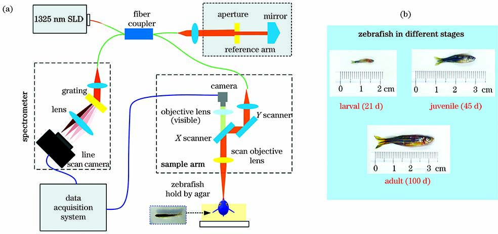
    Fig. 1. OCT system and zebrafish samples. (a) Schematic of OCT setup and photograph of zebrafish hold by agar; (b) photograph of zebrafish samples from three age groups
    OCT in vivo imaging of zebrafish head. (a) Continuous B-scan OCT images; (b) constructed three-dimensional OCT images; (c) virtual segmentation results of three-dimensional OCT images
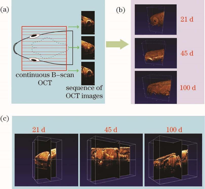
    Fig. 2. OCT in vivo imaging of zebrafish head. (a) Continuous B-scan OCT images; (b) constructed three-dimensional OCT images; (c) virtual segmentation results of three-dimensional OCT images
    Coronal views of images of zebrafish brains from three age groups. (a) OCT images; (b) sectional staining images
    Fig. 3. Coronal views of images of zebrafish brains from three age groups. (a) OCT images; (b) sectional staining images
    Horizontal views of images of zebrafish brains from three age groups. (a) OCT images; (b) sectional staining images
    Fig. 4. Horizontal views of images of zebrafish brains from three age groups. (a) OCT images; (b) sectional staining images
    Measured brain volume of zebrafish. (a) Three dimensional images of zebrafish brains; (b) measured results of brain volume
    Fig. 5. Measured brain volume of zebrafish. (a) Three dimensional images of zebrafish brains; (b) measured results of brain volume
    Brain area of zebrafish obtained based on Fig. 4. (a)--(c) Comparison of sectional staining results and OCT results; (d)--(f) enlarged sectional staining results
    Fig. 6. Brain area of zebrafish obtained based on Fig. 4. (a)--(c) Comparison of sectional staining results and OCT results; (d)--(f) enlarged sectional staining results
    ImagingtechniqueContrast sourceResolutionPenetrationdepthImagingobjectImagingspeedLabelCost
    ConfocalmicroscopyFluorescence<1μm<500μmEmbryoLowYes***
    Light sheetmicroscopyFluorescence1--10μm0.1--5mmEmbryo andinfantFastYes***
    PhotoacoustictomographyOpticalabsorption100μm--1mm<10mmAdultFastNo***
    Micro-CTX-ray attenuation2--3mmNo limitedAdultMediumNo***
    MRIH+ concentration2--3mmNo limitedAdultLowNo****
    OCTOptical scattering1--10μm2--3mmInfant and adultFastNo**
    Table 1. Comparison of in vivo imaging techniques of zebrafish [8, 11, 25-
    Mao Guangjuan, Lin Yanping, Chen Tingru, Zhang Yiqing, Qiu Ting, Lan Yintao, Xiang Xiang, Fu Hongbo, Zhang Jian. OCT in vivo Three-Dimensional Visualization of Zebrafish Brains from Juvenile to Adult[J]. Chinese Journal of Lasers, 2020, 47(12): 1207002
    Download Citation