
Weiwei Tang, Qiannan Jia, Yong Wang, Ding Zhao, Wei Lyu, Wei Yan, Min Qiu. Light-induced vacuum micromotors based on an antimony telluride microplate[J]. Advanced Photonics Nexus, 2022, 1(2): 026005
- Advanced Photonics Nexus
- Vol. 1, Issue 2, 026005 (2022)
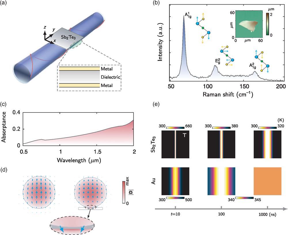
Fig. 1. Optical and thermal properties of hybrid Supplementary Material .
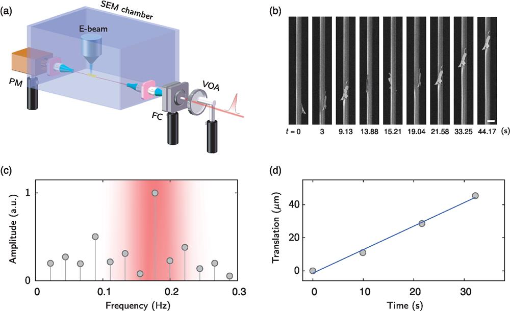
Fig. 2. Experimental observations of a Video 1 , MP4, 4.34 MB [URL: https://doi.org/10.1117/1.APN.1.2.026005.s1 ). (c) Fourier transformation of the detected areas of the
Fig. 3. Manipulating motion speed of Video 2 , MP4, 19.6 MB [URL: https://doi.org/10.1117/1.APN.1.2.026005.s2 ]).
Fig. 4. Stable motion of a Video 3 , MP4, 9.73 MB [URL: https://doi.org/10.1117/1.APN.1.2.026005.s3 ]).
Fig. 5. SEM images of a
Fig. 6. Liquid-like motion of Video 4 , MP4, 895 kB [URL: https://doi.org/10.1117/1.APN.1.2.026005.s4 ]). (b) Zoomed-in microbump [corresponding to the marked rectangle in (a)] showing asymmetric contact angles. Scale bar: Video 5 , MP4, 7.90 MB [URL: https://doi.org/10.1117/1.APN.1.2.026005.s5 ]).

Set citation alerts for the article
Please enter your email address
© Copyright 2018-2021 | Chinese Laser Press. All Rights Reserved 沪ICP备15018463号-20