
Xu Yiwen, Zhang Yunhai, Yang Haomin, Ji Lin, Chang Jian, Liu Chuang, Tang Yuguo. Imaging Technology of Reduced Photobleaching Based on Controllable Light Exposure-Confocal Microscopy[J]. Chinese Journal of Lasers, 2018, 45(4): 407001
- Chinese Journal of Lasers
- Vol. 45, Issue 4, 407001 (2018)
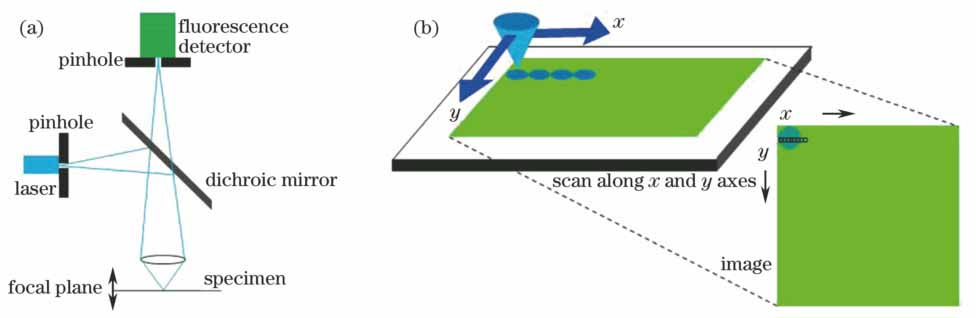
Fig. 1. Schematics of (a) principle of laser spot scanning confocal microscopy and (b) spot scanning process of confocal microscopy
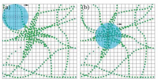
Fig. 2. Spot scanning diagrams of objective pixel. (a) Fluorophore sparse area; (b) fluorophore dense area
Fig. 3. Process of feedback judgment in CLE-CM. (a) Fluorophore with extremely sparse density; (b) fluorophore with high density; (c) fluorophore with medium density
Fig. 4. Schematic of time subsection in upper threshold judgment
Fig. 5. (a) Pixel light-exposure time distribution; (b) real pixel value; (c) reconstructed image of CLE-CM
Fig. 6. Diagram of CLE-CM imaging system
Fig. 7. (a) Standard confocal contrast images; (b) distribution of pixel light-exposure time; (c) distribution of real pixel value; (d) reconstructed images of CLE-CM
Fig. 8. (a) Standard confocal images; (b) reconstructed images of CLE-CM
Fig. 9. Curves of fluorescence intensity

Set citation alerts for the article
Please enter your email address
© Copyright 2018-2021 | Chinese Laser Press. All Rights Reserved 沪ICP备15018463号-20