In the process of medical image acquisition, due to some factors of the image acquisition device (such as improper parameter adjustment and the limitation of the equipment’s inherent attributes) or the conditions of the object itself (that is, the light absorption and reflection of different attributes) makes the signal collecting process and transferring process in the presence of complicated noise model, causing lung CT image has the characteristics of low contrast and visible mask. Therefore, images with poor visual quality seriously interfere with the efficiency of clinical diagnosis and are a significant obstacle to the subsequent use of images. There is a lot of research on medical image enhancement, but the work on lung CT image enhancement is still lacking. Additionally, when processing images, existing image contrast enhancement algorithms based on histogram equalization tend to introduce unnecessary artifacts, produce an artificial appearance, and cause wash-off effects. Therefore, this paper researched lung CT image enhancement.
We devote to overcoming this over-enhancing problem of existing algorithms and then propose an algorithm which can realize appropriate contrast enhancement without introducing new artifacts, that is an image enhancement algorithm based on image segmentation and a total variation model. As is known to all, lung CT images are poor in contrast due to their narrow dynamic grayscale range. And the visual perception of difference relies on gray histogram distribution characteristics to a great extent. Therefore, the research method of contrast enhancement adopted in this article is based on gray histogram transformation. Furthermore, regarding the feature differences between the foreground and background of lung CT images, a segmentation method based on a global threshold is used to segment the lung parenchyma that doctors are interested in for further processing.
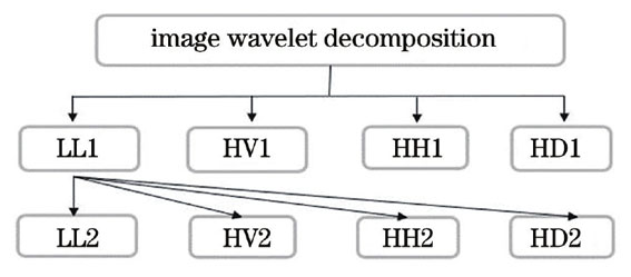
As for the complex noise model in the image, traditional denoising methods are challenging to ensure the regularization of image enhancement results. Consequently, this framework uses the gradient descent method and the total variation framework to separate the image’s noise from the perspective of minimizing energy. Following that, the image structure information and the image detail information will be obtained along with the noise. Then, wavelet transform technique is used to suppress the noise among the image detail information.
The pipeline of the algorithm is as follows, dividing the image into foreground and background firstly, performing bipolar threshold clipping and cumulative distribution function redistribution on the histogram of the foreground lung parenchyma image to form a modified histogram, and then performing Gamma adaptive stretching on the image according to the modified histogram. As a result, the contrast-enhanced foreground image is obtained and fused with the background image as the input of the total variation model. The total variation model then decomposes the image into a texture layer and a structural layer. Next, the texture layer is denoised by the wavelet threshold. Finally, the enhanced image is obtained by fusion of texture layer and structural layer.
This research proposed a framework for improving lung CT images using image segmentation, histogram modification, total variational, and wavelet transform technique (Fig. 2). The subjective analysis of the experimental results shows that the algorithm effectively suppresses the artifacts noise of the image, solves the defect of the existing algorithm over-enhancing lung CT image, comprehensively improves the image contrast, and preserves the complete natural information of the image, as shown in Fig. 3 and Fig. 4. The comparison of image details in Fig. 3 shows that the enhanced image maintains a reasonable degree of regularity in terms of appearance display, texture details, and edge characteristics.
The average value of the objective evaluation index of the experimental results is shown in Table 1. It can be seen that the objective evaluation index parameters of the proposed method have obvious advantages compared with other existing image enhancement methods by comparing the image evaluation index, such as the contrast, grayscale resolution, structural similarity, and absolute mean brightness difference. For instance, the proposed algorithm framework not only fully enhances contrast by increasing grayscale dynamic range display, but also assures the regularity of the enhanced results. The average intensity of the enhanced image by our algorithm is closest to that of the original image, showing the enhanced image has the highest similarity with the original image.
This paper proposes an image enhancement algorithm that solves the issues of low contrast and visible mask in lung CT images. Furthermore, it overcomes the tissues of over enhancement and washout effects which are easy to occur in existing image enhancement algorithms. The research shows that the proposed method can effectively suppress the artifacts noise of the original image in the different areas of the test image, enhance the contrast of the lung CT image, improve the visual effect significantly, and overcome the unwanted artificial artifact greatly. The algorithm is significantly better than other enhancement algorithms in terms of subjective performance evaluation and objective evaluation index. Therefore, the enhancement framework proposed in this paper can provide robust technical support for lung CT image enhancement and improve the efficiency and accuracy of clinical diagnosis and treatment.