• Chinese Optics Letters
  • Vol. 14, Issue 8, 081402 (2016)
Xiumei Liu and Mingli Jiao*
Author Affiliations
  • School of Mechatronic Engineering, China University of Mining and Technology, Xuzhou 221116, China
  • show less
    DOI: 10.3788/COL201614.081402 Cite this Article Set citation alerts
    Xiumei Liu, Mingli Jiao. Behavior of microbubbles on spatially controlled golden nanoparticles[J]. Chinese Optics Letters, 2016, 14(8): 081402 Copy Citation Text show less
    (a) Optical images of the gold nanopillar arrays. (b) Optical image of the gold nanopillar array with the spacing of 1 μm. (c) Scanning electron microscope image of an individual gold nanopillar.
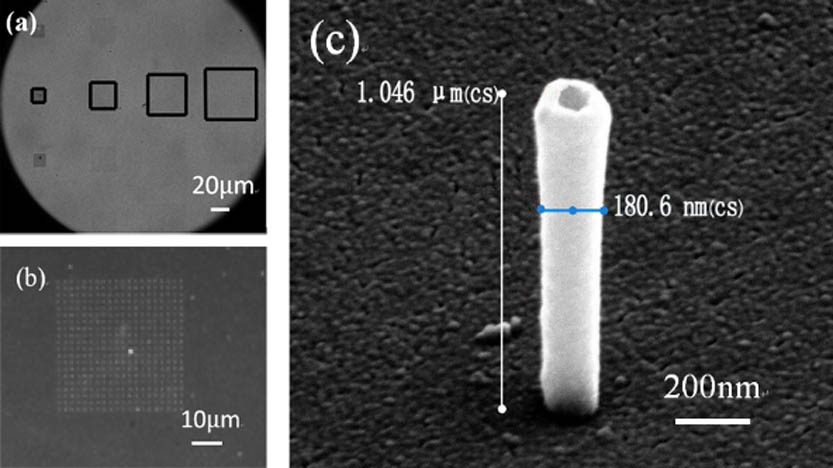
    Fig. 1. (a) Optical images of the gold nanopillar arrays. (b) Optical image of the gold nanopillar array with the spacing of 1 μm. (c) Scanning electron microscope image of an individual gold nanopillar.
    Schematic of the experimental setup.
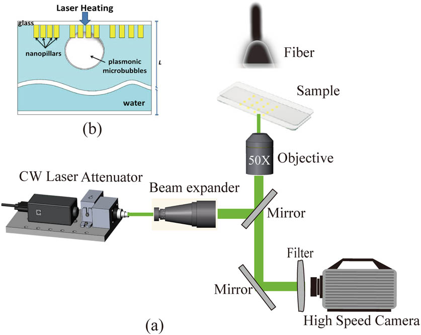
    Fig. 2. Schematic of the experimental setup.
    Sequential images (a)–(h) demonstrate the formation and (i)–(p) show the shrink of a single plasmonic bubble (bottom view) on 1 μm nanopillars. The fluid is degassed water and the absorbed laser power is 12.97 mw.
    Fig. 3. Sequential images (a)–(h) demonstrate the formation and (i)–(p) show the shrink of a single plasmonic bubble (bottom view) on 1 μm nanopillars. The fluid is degassed water and the absorbed laser power is 12.97 mw.
    Plot of the bubble radius versus the time extracted from the top-view images in Fig. 3.
    Fig. 4. Plot of the bubble radius versus the time extracted from the top-view images in Fig. 3.
    Threshold of laser power required for the onset of the plasmonic bubble formation in partially degassed water.
    Fig. 5. Threshold of laser power required for the onset of the plasmonic bubble formation in partially degassed water.
    MB on spatially controlled nanopillars. (a) The images of an MB on different spatially controlled nanopillars at t=100 ms (the error bar is 10 μm). (b) Schematics depicting MB formation due to plasmonic resonance.
    Fig. 6. MB on spatially controlled nanopillars. (a) The images of an MB on different spatially controlled nanopillars at t=100ms (the error bar is 10 μm). (b) Schematics depicting MB formation due to plasmonic resonance.
    Plasmonic bubble on different spatially controlled nanopillars in degassed water.
    Fig. 7. Plasmonic bubble on different spatially controlled nanopillars in degassed water.
    Bubble growth velocity vs. time on different nanopillars.
    Fig. 8. Bubble growth velocity vs. time on different nanopillars.
    Xiumei Liu, Mingli Jiao. Behavior of microbubbles on spatially controlled golden nanoparticles[J]. Chinese Optics Letters, 2016, 14(8): 081402
    Download Citation