Luminescence thermometry can perform noninvasive thermal sensing with high spatial resolution and fast response, emerging as an exciting field of research due to its promising applications in biomedicine. Nevertheless, because of the interaction between light and complex tissues, the reliability and the accuracy of this technique suffer serious interference, which significantly restricts its practical utilization. Here, a strategy to implement effective luminescence nanothermometry is preliminarily proposed by employing the different thermal responses between and energy transfer processes. Different from the traditional ratiometric sensing method, where two luminescence intensities are used as the thermal response parameters, we use two intensity ratios between and near-IR emissions that are obtained under dual excitation as the detecting and reference signals to perform temperature measurement. This multiparameter-based, self-reference thermometry technique, as we define it, exhibits excellent immunity to the influences arising from the fluctuation and loss of pumping sources as well as the luminescence attenuation in media. High thermal sensitivity () and good resolution () are successfully achieved here, accompanied by a measurement error of in a biological environment test, while large errors are observed based on the traditional ratiometric approach (). We believe the viewpoint in this work could boost luminescence thermometry and provide an ingenious route toward high-performance thermal sensing for biological systems.
1. INTRODUCTION
Luminescence thermometry enables accurate, remote, and optically based temperature measurement and has found numerous applications in different fields ranging from scientific research to industrial production [1,2]. However, it is in biomedicine where luminescence thermometry has found the most innovative applications. This thermal sensing technique makes it possible for noninvasive temperature measurement in biological systems, which provides key information for in-depth understanding of biodynamics processes and is of great importance for the early diagnosis of diseases and precise therapies [3–5]. Thanks to the fast development of nanotechnology, thermometry with high spatial resolution can be actualized through diverse luminescent compounds. For instance, organic dyes, quantum dots, nano-diamonds, carbon dots, and rare earth (RE) ion-doped nanocrystals have been widely examined for nanothermometry [6–8]. Based on the temperature-dependent luminescence properties, including emission intensity, spectral band shape, peak position, luminescence lifetime and polarization anisotropy, the thermal information can be extracted through a contactless mode [9–12]. In the past decades, the ratiometric measurement method has emerged as the most used approach to calculate the absolute temperature, where the intensity ratio between two luminescence bands with different thermal responses is defined as the thermosensitive parameter. In this case, the temperature mapping can be easily and simply operated via the spectroscopic approach and high sensing sensitivity also can be guaranteed [13–15]. The ratiometric sensing technique is becoming one of the most appealing thermometric methods, in part because the technique is usually independent of the concentration of thermometers, which helps to improve the measurement accuracy.
Luminescence thermometry with emission bands located within the biological windows (), where the absorption and the scattering of light in biosystems are minimized, enables deep penetration in tissues and has attracted considerable interest in recent years [16–18]. In this sense, the rare earth ion-activated nanothermometers have demonstrated their leading role as probes in biomedicine, not only because of the abundant electron transition channels in the near-IR (NIR) wavelength region but also because of their outstanding advantages of good biocompatibility, low cost, and the simple preparation process [19–22].
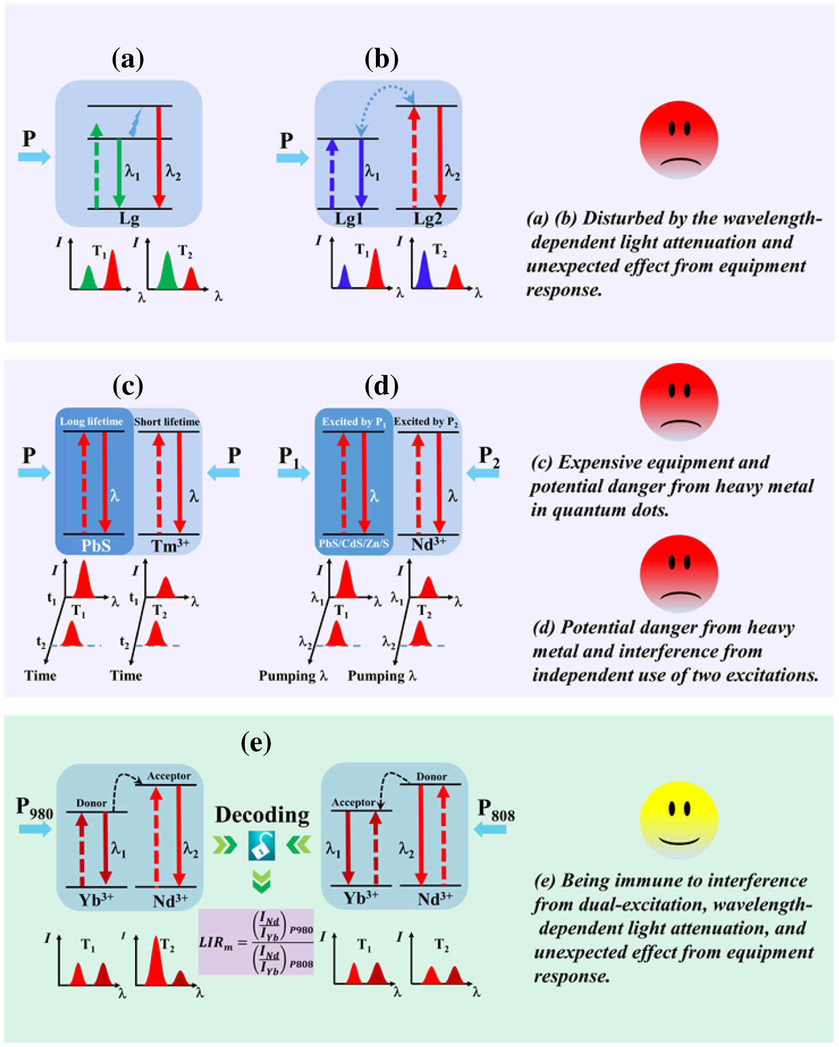
Until now, several schemes have been advanced to conduct temperature sensing based on the luminescence intensity ratio (LIR), and these schemes are summarized in Fig. 1. In the case of traditional thermometry, using thermally coupled levels (TCLs), as shown in Fig. 1(a), for the populations that can be described by Boltzmann-type distribution, the LIR is usually independent of the excitation situation, including the power and the spot area of irradiation on samples because of the intrinsic thermal population between TCLs [23,24]. This unique advantage can effectively avoid an excitation-induced measurement error. Because the limited energy gap between TCLs severely blocks improvement of the measurement sensitivity, ratiometric thermometry using two luminescent groups [Fig. 1(b)] is developed as a flexible way to enhance the thermal sensitivity [25–27]. Since emissions corresponding to these two luminescent groups usually exhibit remarkably different thermal responses, high sensing sensitivities can be achieved. Although the development in luminescence nanothermometry has been witnessed over the past decades, it remains a challenge for the scientific community to develop an effective thermometry strategy suitable for the biological environment. Several questions concerning the reliability of luminescence thermometry are appearing, which are mainly attributed to the interplay between biological components and light. The wavelength-dependent attenuation of light in tissues can lead to the distortion of the spectral structure, making LIR drift away from the actual value. As a result, the thermal readouts acquired by the methods above usually present much large errors [28,29]. Some investigations demonstrated that the wavelength dependent luminescence loss could even result in tens of degrees of measurement error [30,31]. Because of the heterogeneity and the complexity of the biological environment, the precise quantification of light attenuation in tissues is almost impossible [30]. In addition, the reliability of ratiometric thermometry using emissions within different wavelength regions also has been questioned because the luminescence spectra can be affected by other wavelength-associated factors, such as self-absorption of emissions by luminescent centers and the unexpected dependence of the equipment’s response [31,32].
Sign up for Photonics Research TOC. Get the latest issue of Photonics Research delivered right to you!Sign up now
Figure 1.Schematic illustrations of optical temperature sensing based on emissions working in different wavelength regions from (a) thermally coupled levels and (b) two luminescent groups (Lg). Schematic illustrations of optical temperature sensing based on emissions working in identical wavelength regions from (c) two Lgs excited by a single light source and (d) two Lgs excited by two light sources. (e) Dual-excitation decoding multiparameter-based ratiometric thermometry strategy proposed in this work.
One way to break through the limitations in the traditional thermometry technique is to use emissions located within the identical wavelength region. Qiu et al. proposed a ratiometric sensing approach that used two emissions with the same working wavelength (), which were originated from and PbS in nanocomposites [33]. In their work, the time-resolved technique was employed to identify the overlapped NIR emissions from and PbS () quantum dots acquired by a single excitation source, as shown in Fig. 1(c). In this way, the measurement errors arising from the excitation conditions, the selective attenuation of emissions, and the detector response can be suppressed. However, their work required a lot of expensive equipment in the temperature mapping system. Recently, an alternative approach was developed based on the luminescence with same wavelength obtained by different excitations. For example, Long et al. reported optical thermometry based on doped congruent , the red luminescence of which presented different thermal dependences under excitations at 360 and 463 nm [34]. For thermal sensing in the biological environment, Yu et al. constructed a new type of nanocomposites, where two emissions with identical wavelengths from the quantum dots (PbS/CdS/ZnS) and doped fluoride nanocrystals, which were excited by two lasers, were defined as the thermometric parameter [35], as shown in Fig. 1(d). Although the interference of the luminescence attenuation and detector response on thermometric behavior can be avoided by this method, the independent use of two pumping sources would lead to different dependencies of luminescence signals on the pumping conditions. The pumping energy also would be attenuated during the propagation of the light through the tissues and fluids, and it is difficult to know or predict the exact pumping power density upon the detected target. Significant differences between the fixed and the on-target pumping power densities would arise because of the fluctuation and the attenuation of excitation power; consequently, the independent use of different excitation sources has a negative impact on the measurement accuracy [30]. Moreover, the large diameter () of nanocomposites and the heavy metal in the quantum dots used in works further restrict their practical application in biological systems. Therefore, regarding thermal sensing in the biological environment, successful luminescence nanothermometers should not only be small in size and have fine biosafety, but they also should be able to resist interference from the fluctuation and propagation loss of the pumping energy, the luminescence attenuation in the medium, and the different responses of the detector.
Here, we preliminarily propose what we believe, to the best of our knowledge, is a novel temperature sensing strategy that uses the NIR emissions from nanoparticles. Different from the traditional ratiometric sensing method where one luminescence band intensity is defined as the detecting signal and another is used as the reference, the LIR between and that is obtained under dual excitation is employed as the detection and reference signal, as shown in Fig. 1(e). The different thermal dependence of energy transfer (ET) for and within the physiological temperature range enables us to monitor the temperature with high sensitivity and good resolution. More importantly, this multiparameter-based ratiometric thermometry technique, as defined in this work, can effectively avoid interference from excitation and luminescence attenuation, thereby presenting high accuracy and showing great potential for application in in vivo temperature mapping.
2. EXPERIMENT
codoped nanoparticles were prepared through the typical solvothermal approach. The raw materials include (99.99%), (99.998%), (99.999%), oleic acid (90%), 1-octadecene (), NaOH (), (), methanol (), cyclohexane (), and ethanol (). The lanthanide chlorides were dissolved in the mixture of 6 mL oleic acid and 15 mL 1-octadecene with a molar ratio of , , and 1% . The solution was stirred and heated to 100°C for 10 min. The temperature of the mixture was then gradually increased to 160°C and maintained for 30 min, and then cooled to 50°C. After adding 10 mL methanol with (0.1482 g) and NaOH (0.1 g) into the mixture, the reaction was maintained at 50°C for 30 min. Then, the mixture was evaporated at 100°C for 20 min to remove the methanol and water. After that, the reaction mixture was quickly heated to 300°C and maintained for 60 min under argon atmosphere. After the solution was naturally cooled down to room temperature, the centrifugation was performed to obtain the product, which was washed several times with cyclohexane and ethanol to remove impurities. The obtained sample was dried at 80°C in air for 10 h for optical measurement.
The morphology of as-prepared nanoparticles was identified by transmission electron microscopy (TEM) (Tecnai TF20, FEI). The X-ray diffraction (XRD) technique ( diffractometer with Cu radiation, Rigaku) was employed to understand the crystalline phase of sample. A grating spectrometer (, Zolix) equipped with a photomultiplier tube (CR131) and an InGaAs detector (DInGaAs2600-TE, Zolix) was used to measure the luminescence spectra. Two semiconductor lasers, which were centered at 980 and 808 nm, were used as the excitation sources. The temperature-dependent emission spectra were studied using a hot stage (HCP621G, Instec), whose temperature resolution and stability are, respectively, and . The luminescence decay curves were recorded by a digital phosphor oscilloscope (DPO 2024, Tektronix).
3. RESULTS AND DISCUSSION
It is well known that ET can take place for cases where there is an energy mismatch between the donor and the acceptor because this type of nonresonance that commonly exists in the interaction among RE ions can be realized by the assistance of local phonons [36,37]. According to the Miyakava–Dexter theory, the temperature-dependent, phonon-assisted ET rate can be expressed by [38] where is the ET rate at 0 K; is the absolute temperature; is the energy mismatch between donor and acceptor; is the phonon number involved in the ET process; and is Boltzmann constant. Eqs. (1) and (2) describe the ET rates corresponding to processes actualized by emitting and annihilating phonons, respectively. Concerning the ET process between two types of ions (ion A and ion B), the equations above also indicate the different thermal dependence of ET related to and [39,40]. This conclusion also illustrates the different temperature dependence of the luminescence from A or B when they separately serve as donor and acceptor, implying the possibility of ratiometric thermometry by the same emission from one luminescent group. Yet, to our best knowledge, little research has been carried out to exploit the potential value of this mechanism in optical temperature measurement.
Our previous works revealed that temperature has a significant effect on the phonon-assisted ET of as the codoped systems that were excited by a 980 nm laser (i.e., plays the part of the energy donor and acts as the energy acceptor) [41,42]. This mechanism can eventually lead to an obvious enhancement of NIR emissions with an increase in the temperature. Here, to confirm the feasibility of temperature sensing through the diversity of ET between RE ions, the ET of also will be investigated when ions are resonantly excited by an 808 nm laser, namely the case that serves as the donor and serves as the acceptor. Considering the fine biocompatibility as well as the good physical and chemical stability, hexagonal is selected as the host material and : 1% (doping concentrations 1% and 2% represent molar fractions) nanoparticles (NPs) are employed here as the illustrative sample. Figure 2(a) presents the TEM image and the particle size distribution of the as-prepared NPs. The sample shows a uniform spherical shape with an average diameter around 15.8 nm. Figure 2(b) shows the selected-area electron diffraction patterns corresponding to (100), (110), (101), (201), and (002) planes of the hexagonal lattice. The crystal structure also can be confirmed by the XRD pattern. As shown in Fig. 2(c), all of the diffraction peaks in the XRD pattern can be well indexed to the hexagonal-phase with calculated lattice parameters Å and Å, which are in agreement with the reported data in JCPDS #16-0334 (Å, Å). The intense diffraction peaks indicate that the sample is well-crystallized, and the average size determined by Scherrer’s formula is about 16.3 nm, which is close to the result obtained in the TEM image.
Figure 2.(a) TEM image of nanoparticles and the diameter distribution (insert), (b) selected-area electron diffraction pattern, and (c) XRD pattern for the sample.
To fully understand the interaction between and and then explore a promising application in luminescence thermometry, the NIR emissions in NPs that are obtained by 980 and 808 nm laser excitation, respectively, are recorded at temperatures ranging from 20°C to 60°C. As expected, the NIR luminescence in the wavelength region of , which can be attributed to the transition of , manifests dissimilar changing modes as the temperature increases when excited by different lasers, as shown in Figs. 3(a) and 3(c). Thermally enhanced NIR emission is successfully actualized under 980 nm laser pumping and an approximately twofold enhancement has been achieved as the temperature increases from 20°C to 60°C, as shown in the inset of Fig. 3(a). Nevertheless, little variation in luminescence is observed in the case of 808 nm laser excitation. Figures 3(b) and 3(d) illustrate the luminescence mechanism in codoped system irradiated by light sources with different wavelengths. As ions are resonantly excited by 980 nm laser, the ET of is implemented by absorbing phonons because of the energy mismatch between these two types of ions [42,43]. With respect to 808 nm laser excitation, the state of is populated via ground state absorption, the ions upon which relax to the state and then act as a sensitizer to activate through ET of , accompanied by the release of phonons [44,45].
Figure 3.NIR luminescence spectra of and normalized luminescence intensities (inset) at different temperatures when excited by (a) a 980 nm laser and (c) an 808 nm laser, respectively. The photoluminescence mechanism involved in the case of (b) 980 nm excitation and (d) 808 nm excitation, respectively.
If we denote , and as the population densities for , and states, respectively, the photophysical processes above can be described by the these rate equations: under 980 nm laser excitation, under 808 nm laser excitation, where and are the Planck constant and light speed, respectively; denotes the absorption cross-section of at 980 nm (808 nm); , and () are the power, irradiation spot area on the sample, and wavelength for 980 (808) nm laser, respectively; and are the ET coefficients between and ; and are the total decay rates related to and states, respectively; and and are the nonradiative and the radiative decay rates for state of , respectively. From Eqs. (3) and (7), it is clear that under steady-state conditions, the forward ET (FET) process from sensitizer to activator would be more intense against the back ET (BET). Therefore, to comprehend the temperature dependence of NIR luminescence, it should be started from the analysis of FET occurring between and ions.
As an important parameter to evaluate the ET process, the ET rate can be directly calculated by the decay time experiments [46]. The luminescence decay curves for and : in the singly doped and codoped NPs are measured at temperatures ranging from 20°C to 60°C, as presented in Figs. 4(a)–4(d). Then, the ET rates for and as a function of temperature are determined by , where and are the lifetimes of donor ions in the singly doped and donor-acceptor codoped samples, respectively [47,48]. As seen from the results in Figs. 4(e) and 4(f), the ET of excited by 808 nm laser displays a thermal inertness behavior within the physiological temperature range. In contrast, the thermal-induced increment of relevant to ET of is achieved under 980 nm laser excitation, indicating that ET from to is thermally active. The ET efficiencies are also estimated by [48], as shown in Figs. 4(e) and 4(f), the changing tendency of which is similar to that of the ET rate. As a result, the difference in FETs of finally produces different thermal responses of NIR luminescence, which allows us to monitor the temperature through the intensity ratio of luminescence gained with dual excitation. If the NIR luminescence intensity () of acquired by the 980 nm laser excitation is defined as the detection signal and the gained by the 808 nm laser excitation is regarded as the reference signal, the LIR used for temperature mapping can be obtained by solving Eqs. (3)–(7) under steady-state:
Figure 4.Luminescence decay curves and lifetimes for in (a) singly doped and (b) codoped NPs. Luminescence decay curves and lifetimes for in (c) singly doped and (d) codoped NPs. ET rates and efficiencies for (e) and (f) at different temperatures.
Considering that just a few ions can be excited during the ordinary photoluminescence process, and are approximately equal to the doping concentration of the and ions, respectively. Due to the employment of same emission bands, the LIR is successfully free from the influence of selective loss in the luminescence signals and the response of the detector. However, the LIR is closely related to the power and the spot area of the pumping lasers, as shown in Eq. (8). This result illustrates that the measurement error originating from the excitation cannot be avoided. To achieve reliable luminescence thermometry, another reference emission signal that can remove the negative effect of pumping must be introduced.
Here, it is noteworthy that in the codoped system, the NIR-emitted levels of donor and acceptor will enjoy the same number of photon when irradiated by the 980 or 808 nm laser; i.e., a single photon process is involved in the excitation of and , as shown in Figs. 3(b) and 3(d). Therefore, both and luminescence intensities show the same dependence on the excitation. In other words, the influence of the excitation fluctuation on the thermal sensing can be removed by using the luminescence as an additional reference. So, if we define the ratio between and luminescence intensities as the detection and reference signals instead of the simple luminescence intensity of , the independence of thermometry upon excitation can be further actualized. To check this viewpoint, the LIR between , which are separately acquired by the 980 nm [] and the 808 nm [] laser pumping, is deduced from the rate equations above and
As expected, the modified LIR () by luminescence intensity is impervious to the interference originated from the pumping condition and the luminescence attenuation and only correlates with the luminescent properties of RE ions. This unique characteristic especially presents its potential utilization in optical thermometry. To discriminate from the traditional ratiometric sensing method, we define the thermometric approach developed here as multiparameter-based ratiometric thermometry. The applicability of such a thermal sensing strategy is further verified through the experimental scheme presented in Fig. 5(a). The NPs are put in a heating stage that is precisely regulated by a temperature controller. To ensure the same optical path, the outputs of 980 and 808 nm lasers are combined through a fiber combiner. Spectrometer equipped with a photomultiplier tube (PMT) and an InGaAs detector is used to record the NIR emissions from and ions. Two slices of chicken breast () and two cuvettes (10 mm optical path length) containing water are placed perpendicular to the optical axis in the excitation and emission paths to mimic the biological environment.
Figure 5.(a) Schematic of experimental system to imitate thermometry in a biological environment. Luminescence spectra of and normalized luminescence intensities (inset) at different temperatures when excited by: (b) a 980 nm laser and (c) an 808 nm laser, respectively. (d) Plot of versus to calibrate thermal sensing behavior (the pumping power densities corresponding to 980 nm and 808 nm lasers are set at 6.5 and , respectively). (e) Repeatability of measured .
First, the luminescence spectra are measured without tissues and cuvettes to obtain the temperature-sensing calibration curve. The NIR emission spectra and normalized luminescence intensities of , which are measured under excitation by a 980 nm laser and an 808 nm laser, respectively, are shown in Figs. 5(b) and 5(c). According to Eq. (9), the thermometric parameter at different temperatures is acquired using the integrated intensities of emission bands for and , as shown in Fig. 5(d). The thermally-induced changing tendency of can be well depicted by , where and are constants and fitted to be about and , respectively. Good repeatability has been observed, as shown in Fig. 5(e), together with a sensitivity S and a resolution estimated to be about and 0.35°C, respectively. We also prepared containing different concentrations of and . The sensitivities for and are 2.1%, 1.9%, 1.8%, 1.9%, 2.0%, and , respectively, which are smaller than that of . The quantum efficiencies of and NIR emissions used for thermal sensing in this sample are also measured at room temperature, which are estimated to be about 0.3% and 16% under 980 nm laser excitation and about 11% and 9% under 808 nm laser excitation, respectively. Table 1 lists the thermometric properties of reported luminescent thermometers based on the traditional ratiometric method. It can be observed that the thermal sensitivity achieved here is higher than the TCL-based ratiometric methods because of the limited energy gap between TCLs [13,49–51]. In addition, the measurement sensitivities based on the Stark splitting levels are even lower: about and for and ions, respectively [18,52,53]. The obtained sensitivity is also higher than or comparable with the ratiometric sensing approaches actualized by two different luminescent groups [35,43,54–56]. Meanwhile, the present strategy provides a good thermal resolution compared to previous literatures.
Thermometric Properties of Luminescent Thermometersa
Nanothermometers
Sensing parameter
𝛌 (nm)
()
𝛅 (°C)
Ref.
Two intensity ratios (or four intensities)
980/808
2.20
0.35
This work
Two intensities
980
0.94
0.40
[49]
Two intensities
980
1.20
0.40
[50]
Two intensities
977
1.95
[51]
Two intensities
808
0.19
–
[52]
Two intensities
808
0.15
–
[53]
Two intensities
1319
0.75
–
[18]
Two intensities
980
2.32
–
[54]
Two intensities
980
0.70
–
[55]
Two intensities
980
2.60
0.14
[43]
Two intensities
808
0.94
–
[56]
Two intensities
980
1.04
0.30
[13]
Two intensities
322
5.30
–
[57]
TbEu–PDA
Two intensities
377
0.07
–
[58]
Two intensities
690
5.00
0.30
[59]
Two intensities
808/830
1.50
1.80
[35]
and denote the maximum sensitivity and the resolution in the physiological temperature range.
Next, we will verify the advantage of this multiparameter-based thermometric technique in the anti-disturbance ability of pumping. For this purpose, the luminescence spectra of and are measured at 40°C with different excitation powers. Considering the large absorption of water molecules at 980 nm, the 980 nm laser power should be carefully adjusted when applying it to biosystems. Therefore, the excitation powers are kept at relatively small values to minimize the possible influence of a laser on the measurement (980 nm laser: ; 808 nm laser: ). Based on the calibration curve, the relevant temperature values under different excitation powers are calculated, as presented in Fig. 6(a). A slight increase in the temperature with the excitation power is observed, which would be probably due to the laser-induced heating effect. Clearly, although the pumping power changes in a relatively large region, it has little impact on the thermal sensing improved by using luminescence as additional reference signal. Such outstanding thermometric performance can also be demonstrated by comparison with the case where temperature measurement is performed just by the LIR of ions expressed in Eq. (8), where a distinct deviation of tested value from actual temperature is observed, as shown in Fig. 6(b). This result simultaneously reveals the fact that the temperature sensing strategy developed here can guarantee precise temperature measurement in a large dynamic range of pumping power. Such a capability is especially meaningful for a situation where it is necessary to appropriately enhance the excitation power to ensure the SNR of the luminescence.
Figure 6.(a) Influence of pumping powers on the multiparameter-based thermometry strategy under dual-excitation. (b) Influence of the excitation power on LIR-based thermometry.
To confirm the superiority of this multiparameter-based thermometry method in a biosystem, the chicken breast slice and the cuvette filled with water are placed in the optical path to emulate the tissue environment. The temperature of the heating stage is still fixed at 40°C. The experiment is carried out in three cases: Case i, the excitation optical path is inserted with the cuvette and chicken breast slice, where the interference arising from the loss of pumping energy can be known; Case ii, the situation of an emission optical path inserted with the cuvette and chicken breast slice is studied to understand the impact of luminescence attenuation on thermometric behavior; Case iii, both of the excitation and emission optical paths are embedded with the cuvette and chicken breast slice to check the comprehensive effect of tissue environment on the sensing approach. To ensure a good SNR, the excitation powers for the 980 nm laser and the 808 nm laser are set at 320 mW and 80 mW, respectively. Figures 7(a)–7(d) present the luminescence spectra of and recorded in different situations. Not only are the emission intensities weakened, but the spectrum profile also is distorted, which are mainly ascribed to the selective loss of light (pumping laser and luminescence) in water and tissue. However, the exhibits little difference. The calculated temperature values based on are 39.2°C (Case i), 40.6°C (Case ii), and 38.9°C (Case iii), respectively, which is much closer to the actual temperature, as shown in Fig. 7(e). For comparison, the results estimated from the LIR defined in Eq. (8) and the intensity ratio (RNY) between the NIR emissions of and obtained by the 980 nm laser excitation, which can also be used to detect temperature [43], are presented in Fig. 7(e). Obviously, large measurement errors are observed by using LIR and RNY. When using LIR as the thermometric parameter, the impact of wavelength-dependent luminescence attenuation on temperature sensing (Case ii) can be suppressed . However, a large reading error of temperature () is obtained in Case (i) due to the different energy losses of the 980 nm laser and the 808 nm laser in water and tissue, which finally results in different changing degrees in the luminescence. Similarly, although the utilization of RNY can lead to a fine measurement result () in Case i, a false thermal reading () is observed in Case ii because of the wavelength-dependent emission attenuation. In Case iii, both the LIR and the RNY () fail to precisely map the temperature. These results further illustrate the superiority of the multiparameter-based luminescence thermometry technique. Since the ET process between and is closely related to the host matrix and the RE doping concentration, the thermometric performance could be further improved by optimizing the luminescent materials, which will be addressed in our future research.
Figure 7.NIR luminescence from and in acquired by (a), (b) 980 nm laser excitation and (c), (d) 808 nm laser excitation, respectively, in different cases. (e) Temperature values calculated by different thermal-responsive parameters. Case 0 denotes the spectral measurement without the chicken breast slice and the cuvette in the optical path.
In summary, we propose what we believe, to the best of our knowledge, is a new strategy to actualize effective luminescence thermometry in a biological environment. Under the excitation of the 980 nm laser, thermally enhanced NIR luminescence from in codoped nanoparticles is obtained because of the strengthened energy transfer from to as the temperature increases from 20°C to 60°C. In contrast, NIR luminescence is hardly affected by the temperature when pumped by the 808 nm laser due to the thermal inertness of the energy transfer for . Based on the different thermal responses of the energy transfer between the RE ions, ratiometric thermometry through the same emission from ions is actualized. More importantly, to prevent the temperature measurement from being influenced by the dual excitation, the emission of is used as additional reference signal. This multiparameter-based ratiometric method exhibits outstanding properties, including high thermal sensitivity (), fine resolution (0.35°C), and a measurement error of approximately 1.1°C in a biological environment test. Therefore, we believe the present study provides a more reliable and precise thermal sensing method that has promising application in the field of biomedicine.