Lipid droplets are important organelles closely associated with various cellular physiological activities. Confocal fluorescence imaging is a powerful tool for observing lipid droplets and studying their diverse functions. However, lipid droplet fluorescent probes with the high fluorescence intensity and labeling selectivity required for cellular lipid droplet fluorescence imaging are limited, severely limiting the in-depth study of lipid droplets. In this study, we develop Lipi-QL, a quinoline-derivative lipid droplet fluorescent probe with fluorescence-switching properties.
The probe exhibits high selectivity for lipid droplet labeling owing to its sensitive polar quenching fluorescence properties. The donor-type molecular structure also confers high fluorescence intensity and large Stokes shifts on the probe. When using this probe for confocal fluorescence imaging of cellular lipid droplets, significantly better labeling selectivity is achieved at varying concentrations than when using the commercial BODIPY 493/503 lipid droplet probe. Additionally, three-dimensional confocal imaging of fixed cells and four-color confocal imaging of live cells are performed using this fluorescent probe. The development of this probe provides a powerful tool for studying the physiological functions of lipid droplets and provides a new idea for the design of new highly labeled selective fluorescent probes.
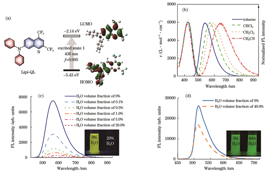
As shown in Fig.1(c), the probe exhibits highly efficient fluorescence emission when the water volume fraction is 0, indicating that it can exhibit high fluorescence intensity within lipid droplets. When the water volume fraction gradually increases, the probe exhibits extremely sensitive fluorescence quenching properties: quenching most of the fluorescence emission when the water volume fraction is only 1%. When the water volume fraction increases to 20%, the probe's emission is almost completely quenched, and the fluorescence signal disappears. This indicates that even if a small portion of the probe enters the cell and stains organelles other than lipid droplets, the fluorescence emission is quenched by the polar environment in which it is placed, thus showing a high selectivity for lipid droplet staining. We also test the fluorescence switching characteristics of the commercial lipid droplet dye, BODIPY 493/503. As shown in Fig.1(d), the fluorescence quenching of BODIPY 493/503 in the dioxane solution with 40% water volume fraction is not apparent, which may be the main reason for its poor lipid droplet staining selectivity. Figure 3 shows that the Lipi-QL fluorescent probe efficiently stains cellular lipid droplets at different concentrations. In contrast, BODIPY 493/503 stains lipid droplets much less selectively, staining other membrane-like cellular structures in addition to cellular lipid droplets with a lower imaging signal-to-noise ratio. This staining selectivity comparison highlights the significant advantage of the polar quenching luminescence property of the Lipi-QL fluorescent probe for the efficient and selective labeling of cellular lipid droplets. After washing the free probe with phosphate buffered saline (PBS), three-dimensional confocal imaging is performed. The experiment is performed at a high xy-plane point resolution with a small z-sweep step (200 nm) to obtain high-quality 3D confocal photographs (Fig.4). The spatial distribution of intracellular lipid droplets can be seen clearly in this photograph, demonstrating the usefulness of the probe for 3D confocal imaging. The Lipi-QL fluorescent probe is also used for multicolor confocal imaging because of its excellent performance. The nuclei, lipid droplets, lysosomes, and mitochondria of live HeLa cells are stained with the Hoechst 33342 commercial dye for nuclei, Lipi-QL commercial dye for lipid droplets, LysoTracker Deep Red commercial dye for lysosomes, and MitoTracker Deep Red commercial dye for mitochondria, respectively. High-quality four-color confocal images of living cells are successfully obtained by performing confocal fluorescence. Based on the different absorption and emission spectra of these four fluorescent probes, imaging is performed through line-by-line scanning, effectively avoiding the occurrence of crosstalk between individual fluorescent channels.
In conclusion, an advanced lipid droplet fluorescent probe with fluorescence switching properties, Lipi-QL, is developed in this study, which allows for the efficient and selective labeling of cellular lipid droplets. The probe also has high fluorescence brightness, a large Stokes shift, and good biocompatibility. Based on these excellent properties, high-quality three-dimensional confocal imaging of fixed cells and four-color confocal imaging of live cells are successfully achieved using this probe, highlighting its utility in lipid droplet fluorescence imaging. The development of this probe provides an effective tool for cell biology studies of lipid droplets and a new approach for the design and synthesis of highly labeled selective fluorescent probes.