Photoacoustic computed tomography (PACT) is an important photoacoustic imaging modality. Compared with photoacoustic microscopy, PACT can detect biological tissues located several centimeters deep without external contrast agents. Equipped with a multi-channel data acquisition card, PACT has the potential for high-speed imaging under a large field of view and is currently used in clinical and preclinical applications, such as whole-body imaging of small animals and human organs. However, skin tissue contains a lot of melanin, and the high-intensity photoacoustic signal from the skin covers the deep subcutaneous tissue information during the imaging process, hindering the en-face display and analysis of the photoacoustic image of the region of interest. Existing works have successfully removed most of the skin signals in photoacoustic images, but there are still some existing problems: (1) most of them are based on photoacoustic microscopic images of shallow tissues or directly extracted vascular structures in the background; the skin removal of deep tissue PACT images has not been reported; (2) the current pixel-level manual labeling takes a lot of time, and there are shortcomings of low extraction accuracy and low efficiency; (3) owing to reconstruction artifacts and changes in light intensity, the signal amplitudes of the skin area are uneven, and there exists many small segments that cannot be distinguished from the background, which increases the difficulty of extracting a complete and continuous skin signal.
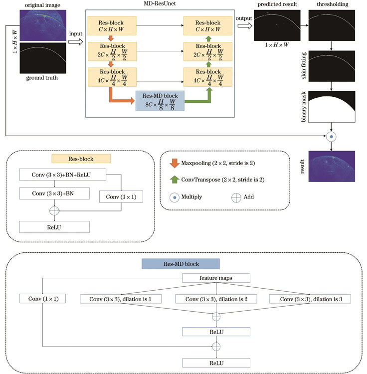
Considering the continuity of the skin tissue and the uniformity of the thickness of the local imaging area, this study proposes a U-shaped deep learning (DL) model that combines multi-scale perception and a residual structure (MD-ResUnet) to automatically remove skin areas in PACT deep tissue photoacoustic images. The introduction of the residual structure in this model can integrate low- and high-level feature information to prevent model degradation, and the multi-scale dilated convolution blocks can increase the continuity and integrity of skin extraction. In the skin segmentation task, a single-type skin region label was proposed as the ground truth, which significantly reduces the complexity of data annotation, compared with the previous pixel-level multi-type annotation. Subsequently, an algorithm of skin integrity fitting and skin mask generation was designed based on the extracted binary image of the skin, to realize the automatic removal of the skin signal in the PACT image. A total of four PACT datasets were used in our experiments, two of which were used for model optimization and two for experimental verification.
The photoacoustic images of the peripheral blood vessels of human legs from PACT verified the correctness and effectiveness of the proposed method on high-precision extraction and removal of skin tissue. In the task of skin segmentation, the comparative experiments with the existing network models of Unet and Res-Unet, show that the DL model MD-ResUnet proposed in this study can fit most of the narrow skin segmentation gaps, effectively shorten the large segmentation gaps, and the extracted skin is overall more accurate, smooth and continuous (Fig. 4). Compared with the existing skin removal works, the deep learning method proposed in this study can thoroughly remove the skin signal and restore a more realistic and clear deep tissue structure (Fig. 5). Quantitative analysis shows that the reconstruction error of the skin-free image has dropped by 50%?70%, and the peak signal-to-noise ratio is averagely increased by 4.5 dB (Table 2), which may provide an effective method for the high-definition display of deep tissue PACT images.
This study proposes a novel skin removal method for PACT deep tissue images with skin region segmentation as the core and designs a new U-shaped DL network MD-ResUnet to achieve the skin segmentation task. The proposed single-class skin-area labeling method significantly reduces the complexity of data processing, and the boundary fitting and mask generation methods realize the complete removal of skin areas, providing an effective method for high-quality deep tissue image generation in PACT. However, the network model proposed in this study cannot yet achieve fully continuous skin-region extraction, and there are still partially disconnected skin gaps. In addition, the experiment in this study is based on the imaging of the peripheral blood vessels of the human leg. The surface of this tissue is relatively regular, and the overall shape of the skin is arc-like, which is convenient for DL to grasp its overall structure features. For imaging tissues with complex surfaces, such as fingers and wrists, the surface shape of the skin is variable, and there will be more significant illumination differences in the same image frame, resulting in increased uneven skin area signals. In the future, we will explore advanced DL network models to implement the extraction of fully continuous skin surfaces in PACT images.