• Chinese Journal of Chemical Physics
  • Vol. 33, Issue 5, 590 (2020)
Juan Ren1, Xian-yi Zhang2, and Xiang-lei Kong1、3、*
Author Affiliations
  • 1State Key Laboratory of Elemento-organic Chemistry, College of Chemistry, Nankai University, Tianjin 300071, China
  • 2School of Physics and Electronic Information, Anhui Normal University, Anhui Normal University, Wuhu 241000, China
  • 3Collaborative Innovation Center of Chemical Science and Engineering, Nankai University, Tianjin 300071, China
  • show less
    DOI: 10.1063/1674-0068/cjcp2006089 Cite this Article
    Juan Ren, Xian-yi Zhang, Xiang-lei Kong. Structure of Protonated Heterodimer of Proline and Phenylalanine: Revealed by Infrared Multiphoton Dissociation Spectroscopy and Theoretical Calculations[J]. Chinese Journal of Chemical Physics, 2020, 33(5): 590 Copy Citation Text show less
    The CID mass spectra of ProPheH+ under two different CID experimental condtions: a) Vp-p= 1.0 V and b) Vp-p= 1.5 V.
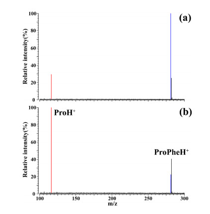
    Fig. 1. The CID mass spectra of ProPheH+ under two different CID experimental condtions: a) Vp-p= 1.0 V and b) Vp-p= 1.5 V.
    (a) ESI mass spectrum of the mixture solution of Pho and Phe, (b) the isolation of the complex ions of ProPheH\begin{document}$ ^+ $\end{document} and (c) IRMPD mass spectrum of ProPheH\begin{document}$ ^+ $\end{document} after a 6 s IR irradiation at 3560 cm\begin{document}$ ^{-1} $\end{document}.
    Fig. 1. (a) ESI mass spectrum of the mixture solution of Pho and Phe, (b) the isolation of the complex ions of ProPheH\begin{document}$ ^+ $\end{document} and (c) IRMPD mass spectrum of ProPheH\begin{document}$ ^+ $\end{document} after a 6 s IR irradiation at 3560 cm\begin{document}$ ^{-1} $\end{document}.
    Relative intensities of fragment and precursor ions of ProPheH+ under different CID conditions.
    Fig. 2. Relative intensities of fragment and precursor ions of ProPheH+ under different CID conditions.
    (a) Experimental IRMPD spectrum of ProPheH\begin{document}$ ^+ $\end{document} in the region of 2700-3700 cm\begin{document}$ ^{-1} $\end{document}, and the calculated IR spectra of (b) PF-ProH-CS-1, (c) PF-PheH-CS-1, (d) PF-PheH-SB-1, and (e) PF-ProH-SB-1.
    Fig. 2. (a) Experimental IRMPD spectrum of ProPheH\begin{document}$ ^+ $\end{document} in the region of 2700-3700 cm\begin{document}$ ^{-1} $\end{document}, and the calculated IR spectra of (b) PF-ProH-CS-1, (c) PF-PheH-CS-1, (d) PF-PheH-SB-1, and (e) PF-ProH-SB-1.
    A view on the 68 optimized structures, corresponding to their energy orders and structural types.
    Fig. 3. A view on the 68 optimized structures, corresponding to their energy orders and structural types.
    The top 20 isomers calculated on the level of M062X/6-311++G (d, p).
    Fig. 3. The top 20 isomers calculated on the level of M062X/6-311++G (d, p).
    Optimized structures of the most stable isomers of ProPheH\begin{document}$ ^+ $\end{document} in four kinds of configurations on the level of M062X/6-311++G(d, p). The H-bonds are identified as dash lines and the lengths are shown in Å.
    Fig. 4. Optimized structures of the most stable isomers of ProPheH\begin{document}$ ^+ $\end{document} in four kinds of configurations on the level of M062X/6-311++G(d, p). The H-bonds are identified as dash lines and the lengths are shown in Å.
    Table 1. PF-ProH-CS-1
    Table 1. Relative energies and free energies (both in kJ/mol) of the four isomers of ProPheH$ ^+ $$ ^{\rm{a}} $.
    Table 2. PF-PheH-CS-1
    Table 2. H-bonds of the four most stable isomers of ProPheH$ ^+ $ shown in FIG. 4. Both PF-PheH-SB-1 and PF-ProH-SB-1 have double intramolecular H-bonds.
    Table 3. PF-PheH-SB-1
    Table 3. The relative energies, free energies at 298K (both in kj/mol) and their calculated ratios at 298K of all 68 isomers calculated on the level of M062X/6-311++G (d, p).
    Table 4. PF-ProH-SB-1
    Juan Ren, Xian-yi Zhang, Xiang-lei Kong. Structure of Protonated Heterodimer of Proline and Phenylalanine: Revealed by Infrared Multiphoton Dissociation Spectroscopy and Theoretical Calculations[J]. Chinese Journal of Chemical Physics, 2020, 33(5): 590
    Download Citation