• Chinese Optics Letters
  • Vol. 15, Issue 11, 111701 (2017)
Xiangwei Lin, Mingjian Sun*, Naizhang Feng, Depeng Hu, and Yi Shen
Author Affiliations
  • Department of Control Science and Engineering, Harbin Institute of Technology, Harbin 150001, China
  • show less
    DOI: 10.3788/COL201715.111701 Cite this Article Set citation alerts
    Xiangwei Lin, Mingjian Sun, Naizhang Feng, Depeng Hu, Yi Shen. Monte Carlo light transport-based blood vessel quantification using linear array photoacoustic tomography[J]. Chinese Optics Letters, 2017, 15(11): 111701 Copy Citation Text show less
    (Color online) MC light transport model for an artificial blood vessel. The target location and beam pattern are at the (a) longitudinal and (d) tangential direction. The corresponding fluence distribution and the absorbed energy density are (b, c) along the x–z slice (y=0 cm) and (e, f) at the y–z slice (x=0 cm).
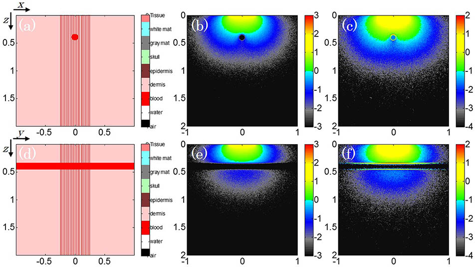
    Fig. 1. (Color online) MC light transport model for an artificial blood vessel. The target location and beam pattern are at the (a) longitudinal and (d) tangential direction. The corresponding fluence distribution and the absorbed energy density are (b, c) along the xz slice (y=0cm) and (e, f) at the yz slice (x=0cm).
    (Color online) Numerical simulation configurations (a) x–z and (b) y–z direction of the PA wave propagation and detection.
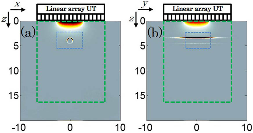
    Fig. 2. (Color online) Numerical simulation configurations (a) xz and (b) yz direction of the PA wave propagation and detection.
    (Color online) Conventional BP reconstructed PA images (a) and (c), and conventional BP reconstructed PA images (b) and (d) with the fluence compensation from the MC light transport model.
    Fig. 3. (Color online) Conventional BP reconstructed PA images (a) and (c), and conventional BP reconstructed PA images (b) and (d) with the fluence compensation from the MC light transport model.
    PAT setup based on the linear array transducer.
    Fig. 4. PAT setup based on the linear array transducer.
    (Color online) Phantom study of an artificial blood vessel. (a) The photograph of the phantom. (b) The fluence map at the axis view. (c) and (d) are the co-registered US and PA images separately recovered from the BP and the fluence-compensated BP method. (e)–(g) are the results in the same order at the lateral view.
    Fig. 5. (Color online) Phantom study of an artificial blood vessel. (a) The photograph of the phantom. (b) The fluence map at the axis view. (c) and (d) are the co-registered US and PA images separately recovered from the BP and the fluence-compensated BP method. (e)–(g) are the results in the same order at the lateral view.
    (Color online) In vivo study of a human forearm vessel. (a) The photograph of the vessel. (b) and (d) are the co-registered US and PA images separately recovered from the BP and the fluence-compensated BP method. (c) The fluence map at the axis view. (e)–(g) are the results in the same order at the lateral view.
    Fig. 6. (Color online) In vivo study of a human forearm vessel. (a) The photograph of the vessel. (b) and (d) are the co-registered US and PA images separately recovered from the BP and the fluence-compensated BP method. (c) The fluence map at the axis view. (e)–(g) are the results in the same order at the lateral view.
    Xiangwei Lin, Mingjian Sun, Naizhang Feng, Depeng Hu, Yi Shen. Monte Carlo light transport-based blood vessel quantification using linear array photoacoustic tomography[J]. Chinese Optics Letters, 2017, 15(11): 111701
    Download Citation