AI Video Guide
  AI Picture Guide
 AI One Sentence
Cervical cancer is one of the most common malignant tumors and poses a serious threat to human health. However, because the onset of cervical cancer is gradual, early and effective screening is crucial. Traditional screening methods rely on manual examinations by pathologists, a process that is time-consuming, labor-intensive, error-prone, and often lacks an adequate number of pathologists for cervical cytology screening, making it challenging to meet the current demands for cervical cancer screening. In recent years, several deep-learning-based methods have been developed for screening abnormal cervical cells. However, because abnormal cervical cells develop from normal cells, they exhibit morphological similarities, making differentiation challenging. Pathologists typically need to reference normal cells in images to accurately distinguish them from abnormal cells. These factors limit the accuracy of abnormal cervical cell screening. This study proposes a Transformer-based approach for abnormal cervical cell screening that leverages the powerful global feature extraction and long-range dependency capabilities of Transformer. This method effectively enhances the detection accuracy of abnormal cervical cells, improving screening efficiency and alleviating the burden on medical professionals.
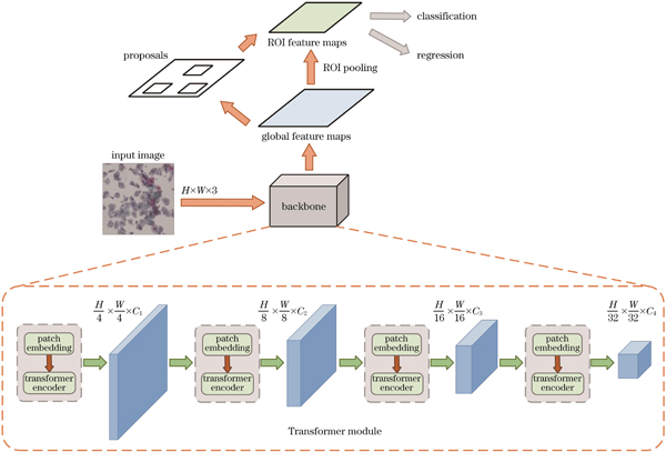
This study introduces a novel Transformer-based method for abnormal cervical cell detection that leverages the powerful global information extraction capabilities of Transformer to mimic the screening process of pathologists. The proposed method incorporates two innovative structures. The first is an improved Transformer encoder, which consists of multiple blocks stacked together. Each block comprises two parts: a multi-head self-attention layer and a feedforward neural network layer. The multi-head self-attention layer captures the correlation of the input data at different levels and scales, enabling the model to better understand the structure of the input sequence. The feedforward neural network layer includes multiple fully connected layers and activation functions and introduces nonlinear transformations to help the model adapt to complex data distributions. We also introduce Depthwise (DW)convolution and Dropout layers to the encoder. DW convolution layer performs convolution operations with separate kernels for each input channel, capturing features within the channels without introducing inter-channel dependencies. Dropout layer reduces the tendency of neural networks to overfit the training data, thereby enhancing the generalization of the model to unseen data. Additionally, we design a dynamic intersection-over-union (IOU) threshold method that adaptively adjusts the IOU threshold. In the initial stages of training, the model can obtain as many effective detections as possible, whereas in later stages, it can filter out most false positive predictions, thereby improving the detection accuracy of the model. Using the proposed method, the model can obtain precise information regarding the location of abnormal cells.
To validate the effectiveness of our proposed method, we compare it with common general-purpose object detection methods. The average accuracy (AP) and AP50 of our method are 26.1% and 46.8%, respectively, surpassing those of all general object detection models (Table 1). In particular, our method outperforms other comparative models by a significant margin in AP metrics, demonstrating that our model not only detects normal-sized targets but can also detect extremely small targets. Additionally, in a comparison with attFPN, a network specifically designed for abnormal cervical cell detection, our method surpasses attFPN in terms of AP by 1.1% (Table 2). Visual inspection of the detection results reveals that our method more accurately identifies target regions with lower false-positive and false-negative rates (Fig.5). Ablation experiments indicate that adopting the improved Transformer encoder method increases AP and AP50 by 1.8% and 2.3%, respectively, compared with the original model. The use of dynamic IOU thresholds results in a 0.6% increase in AP and a 0.9% increase in AP50 compared with the original model (Table 4). Furthermore, a comparison between the dynamic and fixed IOU thresholds in terms of loss and AP during the training process shows that the model with dynamic IOU thresholds experiences a faster loss reduction and achieves a higher AP in the later stages of training (Fig.6).
This study introduces an automatic identification method for abnormal cervical cells utilizing Transformer as the backbone. We further propose an enhanced Transformer encoder structure and a dynamically adjustable IOU threshold. Various comparative experiments on datasets demonstrate that the proposed method outperforms existing approaches in terms of accuracy and other metrics, thereby achieving precise identification of abnormal cervical cells. Through ablation experiments, it is proven that both proposed modules enhance the accuracy of the model in identifying abnormal cervical cells. Overall, the proposed method significantly improves the efficiency of medical image screening, saving medical time and resources, facilitating timely detection of cancerous lesions, and presenting considerable clinical and practical value. Future research may focus on the application of semi-supervised and unsupervised learning in the field of medical imaging to enhance image utilization, improve model detection performance, and better meet clinical requirements.