• Chinese Journal of Lasers
  • Vol. 49, Issue 20, 2007106 (2022)
Wenjing Chen, Zhaohang Jiao, Dongli Qi, Longhai Shen, Nuo Xu, Dongfei Xie, Yifei Li, Jiahua You, Qi Li, and Yu Feng*
Author Affiliations
  • College of Science, Shenyang Ligong University, Shenyang 110158, Liaoning, China
  • show less
    DOI: 10.3788/CJL202249.2007106 Cite this Article Set citation alerts
    Wenjing Chen, Zhaohang Jiao, Dongli Qi, Longhai Shen, Nuo Xu, Dongfei Xie, Yifei Li, Jiahua You, Qi Li, Yu Feng. Diagnosis of Breast Cancer Using Gaussian Function to Fit Autofluorescence Spectrum[J]. Chinese Journal of Lasers, 2022, 49(20): 2007106 Copy Citation Text show less
    Schematic of experimental device
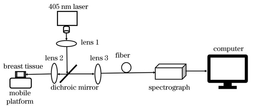
    Fig. 1. Schematic of experimental device
    Fluorescence spectra of slide and stain. (a) Fluorescence spectra of slide; (b) fluorescence spectra of stain
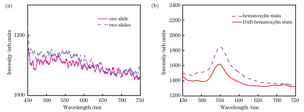
    Fig. 2. Fluorescence spectra of slide and stain. (a) Fluorescence spectra of slide; (b) fluorescence spectra of stain
    Normalized mean fluorescence spectra of breast tissues
    Fig. 3. Normalized mean fluorescence spectra of breast tissues
    Normalized mean fluorescence spectrograms fitted by Gaussian function. (a) Normal breast tissue by SP staining; (b) unstained cancerous breast tissue; (c) cancerous breast tissue by HE staining; (d) cancerous breast tissue by SP staining
    Fig. 4. Normalized mean fluorescence spectrograms fitted by Gaussian function. (a) Normal breast tissue by SP staining; (b) unstained cancerous breast tissue; (c) cancerous breast tissue by HE staining; (d) cancerous breast tissue by SP staining
    NameInstrument information
    LaserWavelength: 405 nm; power: 150 mW
    Dichroic mirrorCritical wavelength: 410 nm; transmission wavelength: >419 nm; transmittance: >80%; reflection wavelength: 360-405 nm; reflectance: >99%
    Fiber probeDiameter: 400 m; wavelength range: 300-800 nm
    Fiber spectrometerSpectral range: 200-1100 nm; scanning speed: 4500 scan·s-1: resolution: 0.8 nm
    Table 1. Experimental instrument information table
    SampleA1A2A3λ1λ2λ3ω1ω2ω3R2
    SP(normal)55.7337.5910.61491.39516.76631.6864.06107.3789.160.998
    Unstained(cancer)19.4872.2331.49492.74520.77630.5939.4182.59147.650.987
    HE(cancer)22.9470.6123.81492.35520.89631.2441.38135.55147.130.992
    SP(cancer)24.4738.5727.86495.14517.25640.8464.3445.05116.200.996
    Table 2. Characteristic parameter information of Gaussian fitting peak
    Breast tissueA517/A492A635/A492
    Normal tissue with SP staining0.67450.1904
    Unstained cancerous tissue3.70791.6105
    Cancerous tissue with HE staining3.07801.0379
    Cancerous tissue with SP staining1.57621.1385
    Table 3. Peak area ratios of A517/A492 and A635/A492 of normal and cancerous breast tissues
    Wenjing Chen, Zhaohang Jiao, Dongli Qi, Longhai Shen, Nuo Xu, Dongfei Xie, Yifei Li, Jiahua You, Qi Li, Yu Feng. Diagnosis of Breast Cancer Using Gaussian Function to Fit Autofluorescence Spectrum[J]. Chinese Journal of Lasers, 2022, 49(20): 2007106
    Download Citation