• Chinese Optics Letters
  • Vol. 17, Issue 12, 123002 (2019)
Chao Shen1、2、*, Nanjing Zhao1、2, Jaka Pribošek3, Mingjun Ma1、2, Li Fang1、2, Xingjiu Huang4, and Yujun Zhang5、**
Author Affiliations
  • 1Key Laboratory of Environmental Optics & Technology, Anhui Institute of Optics and Fine Mechanics, Chinese Academy of Sciences, Hefei 230031, China
  • 2Key Laboratory of Optical Monitoring Technology for Environment, Hefei 230031, China
  • 3Carinthian Tech Research, 9524 Villach, Austria
  • 4Institute of Intelligent Machines, Chinese Academy of Sciences, Hefei 230031, China
  • 5State Key Laboratory of Pulsed Power Laser Technology, National University of Defense Technology, Hefei 230037, China
  • show less
    DOI: 10.3788/COL201917.123002 Cite this Article Set citation alerts
    Chao Shen, Nanjing Zhao, Jaka Pribošek, Mingjun Ma, Li Fang, Xingjiu Huang, Yujun Zhang. Characteristics of optical emission during laser-induced damage events of fused silica[J]. Chinese Optics Letters, 2019, 17(12): 123002 Copy Citation Text show less
    Schematic of experimental setup. The inset is the image of air plasma. The red dashed line is the position of the sample surface, and the white arrow indicates the direction of the laser pulse. The arrangements of the camera and spectrometer are set as an example of the exit surface damage. HWP, half-wavelength plate; PBS, polarized beam splitter; BS, beam splitter; EM, energy meter; FL, focus lens; FC, fiber collector; MO, microscope objective; TL, tube lens.
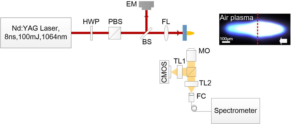
    Fig. 1. Schematic of experimental setup. The inset is the image of air plasma. The red dashed line is the position of the sample surface, and the white arrow indicates the direction of the laser pulse. The arrangements of the camera and spectrometer are set as an example of the exit surface damage. HWP, half-wavelength plate; PBS, polarized beam splitter; BS, beam splitter; EM, energy meter; FL, focus lens; FC, fiber collector; MO, microscope objective; TL, tube lens.
    Typical images of LPP on (a) the exit surface and (b) the input surface. Scanning electron microscope (SEM) morphologies of damage sites on (c) the exit surface and (d) the input surface.
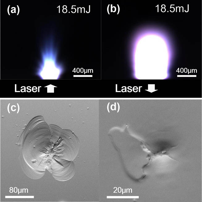
    Fig. 2. Typical images of LPP on (a) the exit surface and (b) the input surface. Scanning electron microscope (SEM) morphologies of damage sites on (c) the exit surface and (d) the input surface.
    (a) Evolution of plasma size with laser energy on the input and exit surfaces, and (b) the ratio between the driving energy on both surfaces deduced by Sedov–Taylor scaling.
    Fig. 3. (a) Evolution of plasma size with laser energy on the input and exit surfaces, and (b) the ratio between the driving energy on both surfaces deduced by Sedov–Taylor scaling.
    Typical plasma emission spectra of bulk damage, exit surface damage, input surface damage, and air plasma from top to bottom. Lines superimposed on the bulk spectra are the fitted line and three Gaussian components. The wavelength range of the inset in the input surface spectra is 572–610 nm. The red arrow indicates the wavelength of 589.25 nm.
    Fig. 4. Typical plasma emission spectra of bulk damage, exit surface damage, input surface damage, and air plasma from top to bottom. Lines superimposed on the bulk spectra are the fitted line and three Gaussian components. The wavelength range of the inset in the input surface spectra is 572–610 nm. The red arrow indicates the wavelength of 589.25 nm.
    Evaluated electron density and excitation temperature of exit and input surface LPPs.
    Fig. 5. Evaluated electron density and excitation temperature of exit and input surface LPPs.
    Speciesλji (nm)Aji (107s1)gEj (eV)
    O I777.3833.691510.74
    844.6463.22910.99
    926.3152.474512.08
    Table 1. Spectroscopic Data Used for Temperature Calculation
    Chao Shen, Nanjing Zhao, Jaka Pribošek, Mingjun Ma, Li Fang, Xingjiu Huang, Yujun Zhang. Characteristics of optical emission during laser-induced damage events of fused silica[J]. Chinese Optics Letters, 2019, 17(12): 123002
    Download Citation