
Kaiqiang Cao, Long Chen, Ke Cheng, Zhenrong Sun, Tianqing Jia. Regular uniform large-area subwavelength nanogratings fabricated by the interference of two femtosecond laser beams via cylindrical lens[J]. Chinese Optics Letters, 2020, 18(9): 093201
- Chinese Optics Letters
- Vol. 18, Issue 9, 093201 (2020)
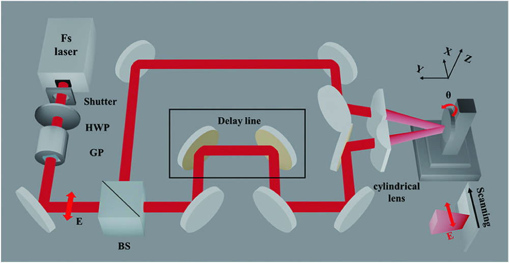
Fig. 1. The experimental setup of femtosecond laser interference via two cylindrical lenses. HWP is half-wave plate, GP is Glan prism, and BS is beam splitter. The double arrow E represents laser polarization.
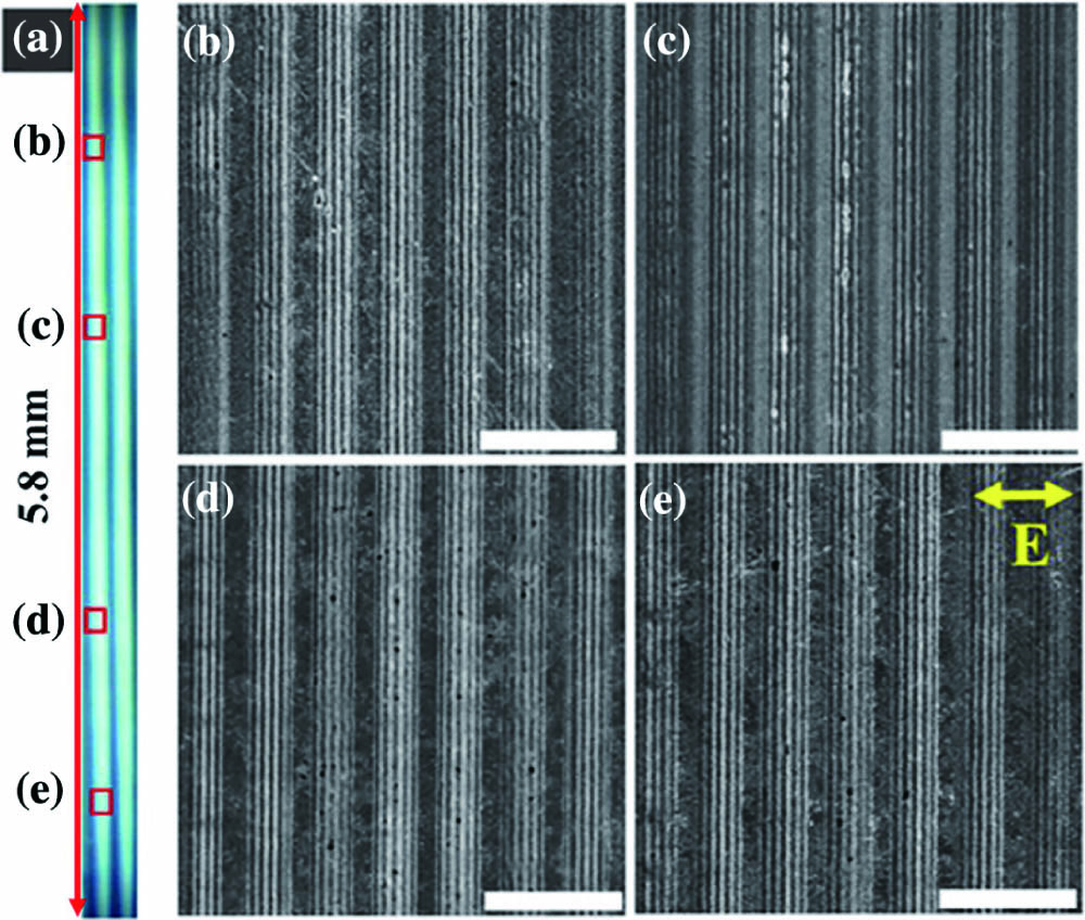
Fig. 2. Microstructures in the ablation area after radiation of three laser pulses. (b)–(e) Enlarged SEM pictures of the areas in squares in (a). The double arrow in (e) represents laser polarization. Laser fluence of single beam is
Fig. 3. (a) Subwavelength periodic ripples by direct writing with a single laser beam. (b) RUSNGs ripples fabricated by direct writing of two-beam interference. (c) RUSNGs ripples without etching in HF solution. The double arrow E represents laser polarization, and the narrow ellipse represents the laser focus.
Fig. 4. Different types of micro/nanostructures depending on the laser fluence and scanning velocity.
Fig. 5. Periods of subwavelength ripples as a function of scanning velocity induced by a single laser beam and two-beam interference.
Fig. 6. (a) Optical image of the colored surface and (b) SEM image of RUSNGs. The scale bar is 5 μm.
Fig. 7. Optical characterization measurements of the periodic ripples. (I) Diagrammatic sketch of the diffraction spectra test; diffraction spectra of (a) subwavelength ripples generated by single laser beam and (b) RUSNGs.
Fig. 8. CIE
Fig. 9. Colorful optical image of the pattern of “raining petals” covered with RUSNGs.
Fig. 10. Colorful optical images of two flowers with nanogratings in different directions. (I) Schematic of the processing method. Flowers with nanogratings in (a) azimuth direction and (b) radial direction. The two enlarged SEM pictures represent the nanogratings in the corresponding squares. The scale bars are 5 μm.

Set citation alerts for the article
Please enter your email address
© Copyright 2018-2021 | Chinese Laser Press. All Rights Reserved 沪ICP备15018463号-20