National Key Laboratory of Photoelectric Technology and Functional Materials (Culture Base) in Shaanxi Province, National Photoelectric Technology and Functional Materials & Application of Science and Technology International Cooperation Base, Institute of Photonics & Photon-Technology, Northwest University, Xi’an 710069, China
A multifunctional photo-thermal therapeutic nano-platform (YR-Si-) was designed through a core–shell structure, expressing the function of bio-tissue imaging, real-time temperature detection, and photo-thermal therapy under 808 nm light excitation. In this system, the core (YR) takes the responsibility of emitting optical information and monitoring temperature, while the shell nano-particles carry most of the photo-thermal conversion function. The temperature sensing characteristic was achieved by the fluorescence intensity ratio using the thermally coupled energy levels (TCLs) of , and its higher accuracy for real-time temperature measurement in the bio-tissue than that of an infrared thermal camera was also proved by sub-tissue experiments. Furthermore, the photo-thermal effect of the present nano-system was confirmed by Escherichia coli (E. coli) and Staphylococcus aureus (S. aureus) ablation. Results indicate that YR-Si- has application prospect in temperature-controlled photo-thermal treatment and imaging in bio-tissues.
1. INTRODUCTION
Photo-thermal therapy (PTT) is a promising alternative reduced invasiveness method for tumor treatment in comparison with traditional chemotherapy and surgery, which uses efficient photo-thermal agents (PTAs) to convert light into heat to ablate cancer cells. For PTT, PTAs determine its success or failure. An ideal PTA not only provides an outstanding light-to-heat conversion performance under the excitation of biological window (BW) region light with deeper tissue penetration depth, but also has excellent bio-security and appropriate size [1–5]. As typical traditional PTAs, nano-sized gold, layered carbon-based compounds, and transitional metal dichalcogenides have been widely used in PTT owing to their strong absorption in the BW region and effective photo-thermal effects [6,7]. However, the nature of nano-material mediated PTT is hyperthermia treatment tumorigenic cells, in which over-high temperature will damage the normal cells and tissues surrounding the diseased tissue while insufficient heat will lead to non-effective therapeutic effects due to the heat production not being effectively monitored and controlled [8–11]. Therefore, it is necessary to develop a nano-platform with a temperature monitor and light-to-heat conversion for application in photo-thermal therapy.
As a newly developed temperature detection method based on the fluorescence intensity ratio (FIR) of the thermally coupled energy levels (TCLs) emissions, it not only offers fast response and high precision but also detects the temperature of internal tissue. However, a traditional temperature monitor using an infrared thermal imaging camera no longer meets the requirement of tissue temperature detection because only the surface temperature could be probed [12,13]. Generally, two emission bands from the TCLs should be fully separated and their energy gap better be below , such as ; ; ; and [14]. Among them, co-doped upconversion nano-particles are extensively used as temperature sensors due to appropriate TCLs of and effective energy migration from to [15–17]. However, the optimal absorption wavelength of centered at 980 nm is consistent with that of molecular water, which seriously limits their applications in biological tissues [18–20]. In order to solve this problem, ions were introduced as a sensitizer to achieve energy transfer of due to their broad absorption at 808 nm in the BW area and over 80% energy transfer efficiency from to [21]. Moreover, the emissions from 850 to 1200 nm of and are suitable for bio-fluorescence imaging under 808 nm. According to previous publications [22–25], the thermal effect from the non-radiative transition of the upconversion (UC) process is not enough to kill cancer cells; it is necessary to introduce effective PTAs in the optical system to enhance photo-thermal conversion efficiency [26–30]. Hence, a new multifunctional nano-platform with the functions of real-time temperature sensing, near-infrared (NIR) imaging, and light-to-heat conversion was designed and achieved through a core–shell structure.
Here, a spherical upconversion nano-particle (YR) was prepared to be used as a temperature monitor and NIR imaging probe. Ultra-small copper sulfide particles were selected as PTAs due to their excellent stability and low cytotoxicity [31]. The multifunctional PTT nano-platform was constructed through combining sphere with nano-particles. In order to increase the water solubility of the nano-particles and the attachment site of the PTAs, as an intermediary was coated on the surface of UC nano-particles. The functions of NIR imaging and temperature sensing for the present system in biological tissues were illustrated by comparative experiments, and its photo-thermal ablation function was also demonstrated by the ablation of bacteria. Results indicate that the current strategy is a promising method to develop an integrated multifunctional photo-thermal therapeutic nano-platform.
Sign up for Photonics Research TOC. Get the latest issue of Photonics Research delivered right to you!Sign up now
2. EXPERIMENT
A. Reagents and Materials
High-purity , , , (99.99%) and analytical grade reagents nitric acid (), urea, ammonium hydroxide (), sodium citrate (), tetraethoxysilane (TEOS), sodium sulfide (), aminopropyltrimethoxysilane (APTMS), and were used as raw materials. In addition, phosphate buffered saline (PBS), Escherichia coli (E. coli) (reference number: ATCC25932), Staphylococcus aureus (S. aureus) (reference number: ATCC6538), nutrient broth, agar medium, and the fresh pork tissue were also used.
B. Synthesis and Processes
1. Synthesis of Nano-Particles
Spherical nano-particles were synthesized via a co-precipitation technique, as described in Ref. [21]. For the representative procedures of preparing about 1 mmol samples, a stoichiometric amount of high-purity lanthanide oxides (∶∶∶) was added in 15 mL nitric acid under stirring and heating until the clarified rare-earth nitrate solution A was obtained. Meanwhile urea (12 g) was added in 50 mL de-ionized (DI) water to obtain solution B; after that, solutions A and B were well mixed and kept at 368 K for stirring 30 min. And then, the precursors were obtained by centrifugating at 10,000 r/min and washing with water. Finally, the final samples nano-spheres were obtained through heating the dried precursors at 973 K for 3 h with a 3 K/min heating rate.
In order to increase the biocompatibility of the samples, the core–shell structured YR@SiO2 (YR-Si) nano-spheres were fabricated via a modified Stöber process [32]. 0.20 g of as-obtained was dispersed in a mixed solution with 72 mL ethanol, 18 mL DI water, and 1.0 mL of concentrated ammonia aqueous solution (25 wt. %) in a 150 mL flask. The resultant mixed solution was stirred for 10 min; then 0.12 mL of TEOS was added to the solution drop by drop. After reaction for 6 h, the particles were washed several times with ethanol and DI water, centrifugally separated with 10,000 r/min, and then dried at 343 K to obtain electronegative YR-Si.
2. Synthesis of Nano-Particles
To connect electronegative ultra-small particles with electronegative YR-Si by electrostatic adsorption, the as-prepared YR-Si nano-spheres (0.2 g) were first dispersed in 100 mL of ethanol and then APTMS (0.35 mL) was added to the solution drop by drop and stirred for 12 h to get the positive YR-Si. Next, the electropositive was collected by centrifugation, washed, and dispersed in 10 mL of DI water.
nano-particles were prepared on the basis of Ref. [33], in which 0.014 g and 0.02 g sodium citrate were added to 100 mL DI water with continuous stirring, and then 2 mL of (0.04 mol/L) was added into the above solution. After 10 min, the brown solution reacted at 363 K for 30 min in the water bath until a dark-green solution was obtained. Following that, the solution was moved to ice-cold water for subsequent use. After that, 0.02 g was dispersed in 60 mL DI water, and the 30 mL solution was then dropped in the above solution and constantly stirred for 1 h to get (YR-Si-) by centrifugation.
3. Photo-Thermal Combined Killing of Bacteria
The photo-thermal effects of samples were detected through thermal ablation of the micro-organism. Bacteria E. coli and S. aureus were transferred to 100 mL sterile nutrient broth solution to get dispersed solution C; D is the PBS buffer solution with (1.0 mg/mL). 50 μL C and 100 μL D were adequately mingled, and the acquired solution was exposed to a 808 nm laser with for 3 min. After that, 20 μL laser treated solution was transferred to the nutrient agar medium and disposed by the spread plate method. After being cultured for 24 h at 310 K, the bacteria colony number was calculated.
C. Characterization
The crystal structures and phase purities were characterized through powder X-ray diffraction (XRD) by a Rigaku-Dmax 3C powder diffractometer (Rigaku Corp, Tokyo, Japan) in the range of . The emission spectra were collected by a spectrophotometer (FLS920, 808 nm semiconductor laser). The morphology and microstructure of the samples were recorded by a transmission electron microscope (TEM, JEM-2100F, Japan) and high-resolution (HR) TEM equipped with an energy dispersive X-ray spectroscope operating at 200 kV. The AXIS-ULTRA DLD (The United Kingdom) with an Al Kα X-ray source was used for X-ray photoelectron spectroscopy (XPS) analysis under analyzer pass energy of 20 eV. The photo-thermal behaviors of samples were received by an InfReC R500 infrared thermal camera (NEC, Japan) and the UV-vis-NIR absorption spectra were obtained by Cary 5000 UV-Vis-NIR by using a Pb smart detector.
3. RESULTS AND DISCUSSIONS
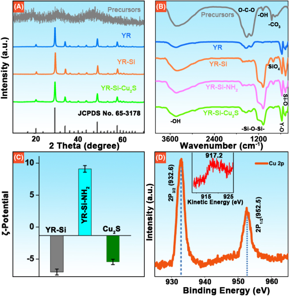
The multifunctional therapeutic nano-system YR-Si- was prepared through four steps. The first step is the synthesis of precursors by the co-precipitation method; the second is to obtain the YR through heating at 973 K. The third is to coat the layer on the YR surface and get the core–shell structure YR-Si, and subsequently obtain the final samples through absorbing nano-particles on the surface of the YR-Si by electrostatic force. The XRD patterns of products at each step are displayed in Fig. 1(A). No peaks were found in precursors, but the diffraction peaks from appeared after calcinating for 3 h at 973 K and matched well with those of the standard (JCPDS No. 65-3178) profile. Results indicated that the precursors were amorphous and the introduction of , , and dopants in the host did not cause the appearance of any impurities. However, the diffraction peaks of the coating layer and in the samples YR-Si and YR-Si- were not observed, which may be due to the amorphous structure of and the low amount of nano-particles.
Figure 1.(A) XRD patterns of precursors, YR, YR-Si, and YR-Si-; (B) FTIR spectra of each step of the synthesized samples of precursor, YR, YR-Si, YR-Si-, and YR-Si-; (C) Zeta potential of YR-Si, YR-Si-, and ; (D) XPS profile of Cu 2p of films.
To further analyze the composition of products in different synthesis steps, their Fourier transform infrared (FTIR) spectra are displayed in Fig. 1(B). The peak at about from O-H of water was found in each step product. Furthermore, O-C-O, -OH, and vibration peaks at , , and were also found in the FTIR spectrum of precursors, which implied that the precursor was . With the formation of after calcinations at 973 K for 3 h, the vibration peaks -OH receded and disappeared with the presence of Y-O peak at . The vibration peaks at 1072 and 464 cm−1 from -Si-O-Si- and Si-O appeared in the FTIR spectra of YR-Si, YR-Si-, and YR-Si-, which declared the formation of through hydrolysis reaction of TEOS.
The final target products YR-Si- were obtained through connecting the third step products YR-Si with , in which the positive charge YR-Si- was obtained through mixing APTMS with YR-Si in ethanol solution and the positive charge attracted the negative-charged nano-particles on the surface of YR-Si by electrostatic interaction. The potential diagrams of YR-Si, YR-Si-, and are shown in Fig. 1(C), which indicates that they have a negative, positive, and negative charge, respectively [23]. The valence state of copper was further explored using XPS analysis. The peaks of Cu and Cu binding energy centered at 932.6 and 952.5 eV are displayed in Fig. 1(D), and the mono-valent species characteristic kinetic energy spectrum peaked at 917.2 eV is presented in the inset of Fig. 1(D) [34,35]. To further confirm the components and microstructure of the intermediate products, the TEM images and elements mapping of YR, YR-Si, and the final products YR-Si- are displayed in Figs. 2(A)–2(I), and the second step product YR maintained the original spherical morphology except in diameter from about 130 to 140 nm [Fig. 2(B)]. For the third step products, a layer of uniform with about 6 nm thickness was coated on the surface of the second product YR to form YR-Si via a modified Stöber process using TEOS as raw materials, as shown in Fig. 2(C). The TEM image of the final product particles YR-Si- is presented in Figs. 2(D) and 2(E); small black particles could be clearly seen on the surface of YR-Si. The HR-TEM images of the core YR and the adsorbed black particles corresponding to the f and g zones in the TEM image of YR-Si- [Fig. 2(E)] are magnified and shown in Figs. 2(F) and 2(G), respectively. It is observed that the lattice distance corresponds to the (222) plane (0.3057 nm) of the cubic (JCPDS No. 65-3178) [Fig. 2(F)], while the lattice distance of about 0.46 nm corresponds to the (211) plane lattice distance of the (JCPDS No. 33-0490) [Fig. 2(G)]. These results confirmed the small particles were and the core was nano-sphere in the final sample particles. Moreover, the core–shell structure of YR-Si- was proved by high-angle annular dark-field scanning TEM (HAADF-STEM) and the cross-section compositional line spectrum shows the distribution characteristics of elements in the final sample YR-Si- core–shell structure [Fig. 2(H)]. Obviously, the elements of Si, Cu, and S are distributed externally and Nd, Yb, Er, and Y are distributed inside YR-Si-. In the meanwhile, the elements mapping images of the samples are also shown in Fig. 2(I), which further confirm the sample is made of Nd, Yb, Er, Y, O, Si, Cu, and S elements. The above results illustrate the final core–shell structured sample YR-Si- was successfully prepared.
Figure 2.(A) Schematic diagram of YR-Si-; TEM images of (B) YR, (C) YR-Si, and (D) YR-Si-; (E) single particle of YR-Si- and high magnification of different zones of f and g are shown in (F) and (G), respectively. (H) HAADF-STEM image and cross-section compositional line profiles of samples and elemental mapping images in (I).
The samples YR, YR-Si, and YR-Si- exhibit similar NIR emissions ranging from 850 to 1200 nm under the excitation of 808 nm light [Fig. 3(A)]. The NIR emission peaks include the characteristic emissions of () at 975 nm and two parts. Comparing the emissions of three samples, it is observed that their photoluminescence (PL) intensities gradually decreased in the order of , which is mainly due to the absorbency of the shell layer or to excitation and emission light [15]. The vis-NIR absorption spectra are provided in Fig. 3(B). No distinct absorption peaks were found in PBS solution but an absorption band from 550 to 1200 nm was observed for , which leads to the absorption of the sample YR-Si- enhanced in comparison with that of YR-Si. This result further indicated that the absorption of to 808 nm excitation light and the vis-NIR emission light from core YR are mostly responsible for the decrease of PL intensity for the sample YR-Si-.
In order to monitor the penetration depth of NIR emitting light from the YR-Si- nano-platform, the normalized depth-dependent integrated intensities of YR-Si- in the NIR region are given in Fig. 3(D). The measurement schematic diagram is displayed in Fig. 3(C). It is found that the PL intensities of NIR emission decreased with the increasing injection depth, but emission light could not disappear until the injection depth was up to 7 mm. Considering that absorption of the molecule of water at 980 nm in biological tissues, Fig. 3(E) shows the integrated intensity ratio () of emission (965–985 nm) and (900–1150 nm) with different injection depths in pork tissues, and the constantly decreased values of R illuminate that the absorption of 980 nm is stronger than that of 900–1150 nm in biological tissues. These results further give the reason for choosing an 808 nm ( sensitizer) instead of a 980 nm NIR laser as the light source, which also confirm the advantages for its potential application in biological imaging.
Figure 3.(A) NIR emission spectra of YR, YR-Si, and YR-Si- under 808 nm; (B) UV-vis-NIR absorption spectra of PBS buffer solution, , YR-Si, and YR-Si- dispersed in PBS; (C) schematic diagram of the NIR light penetration depth in different thicknesses of pork tissue; (D) measured NIR emission intensity as a function of injection depth in pork under 808 nm; and (E) NIR emission intensity ratio of in YR-Si- with different injection depth.
As mentioned in the introduction part, the real-time temperature detection of the focal tumor zone is important for its accurate therapy using a non-contact smart upconversion optical thermometer based on FIR. Here, the typical emission levels and of ions were chosen as TCLs in the nano-system YR-Si-. Sensitivity is one of the most important parameters for the thermometer, which is defined as the rate of the FIR (R) changes with temperature. The absolute sensitivity (), relative sensitivity (), and their FIRs can be demonstrated in the following equations [36]: where and are UC intensities of transitions and , respectively. is the number of ions, is the emission cross section, is the degeneracy, is the angular frequency of the fluorescence transition from to , and and are the Boltzmann and proportionality constants, respectively. is the energy gap between two TCLs of ( and ), and is the absolute temperature. To determine the sensitivity, the temperature-dependent UC emission spectra of YR-Si under 808 nm are shown in Fig. 4(C) in the range of 293 to 493 K. With the normalized emission of (526 nm), the FIR of UC emission from (526 nm) to (549 nm) gradually increases with increasing temperature, which indicates that the population of high-level is growing with the rising temperature. The sensitivity as a function of temperature is calculated and plotted in Fig. 4(D), and the values of YR-Si achieved to the maximum 1.2% at 293 K or at . Those are close to the optimal values 1.32% and in the reported , Er nano-particles [37,38].
Figure 4.Power-dependent temperature of (A) YR, YR-Si, YR-Si- and (B) Y-Si- as a function of time under 808 nm; (C) normalized UC emission spectra of Y-Si at about 537 nm with the increasing temperature to 420 K; (D) absolute/relative sensitivities of Y-Si at different temperatures; and (E) schematic of temperature measurement using FIR and thermal camera. The inset in (E) is surface and sub-tissue temperatures with different 808 nm powers.
Generally, the low quantum efficiency of UC phosphor comes from the high possibility of non-radiation, among which most of energy was transformed to heat. In order to investigate the photo-thermal conversion effect of different samples, the time-dependent temperatures of YR, YR-Si, and YR-Si- were recorded using a thermal camera with the irradiation of 808 nm laser. As shown in Fig. 4(A), the temperature of YR-Si- was much higher than those of YR and YR-Si at each time point under the excitation of the 808 nm laser with a power density. As the exposure time reaches 27 s, the sample temperature grows up to around 299 K for YR or YR-Si, but quickly reaches about 316 K for YR-Si-. With the increasing power density from 0.1 to , the temperature of YR-Si- increases to 373 K and the corresponding thermal images are given for illustration in Figs. 4(A) and 4(B). These results indicate that the sample YR-Si- has the most excellent light-to-heat conversion function.
In order to prove the application of YR-Si- for the measurement of subcutaneous temperature, an appropriate in vivo experiment was designed [as shown in Fig. 4(E)] through comparing the detected temperatures using the FIR technology with the recorded temperature by the infrared camera, in which the PBS solution containing samples of YR-Si- was injected into the pork muscle tissue. The schematic diagram of the experiment is given in Fig. 4(E). The internal temperature of the sub-tissue around the sample particles was detected by the FIR of the present samples, and the surface temperature of tissue was detected by the infrared thermal camera with different power densities under 808 nm [in the inset of Fig. 4(E)]. It can be seen that the detected internal sub-tissue temperature by the FIR technology was much higher and faster than that detected by the thermal infrared camera, which was due to the heat dissipation in the transfer process from the YR-Si- to the pork muscle tissue surface. These results indicate that internal sub-tissue temperature could be accurately monitored in real time using FIR, which implied that FIR has great potential application in detecting the temperature in bio-tissue.
To further prove the YR-Si- photo-thermal effects of the ablation on bacteria (E. coli and S. aureus), the bacteria solutions were incubated for 3 min in PBS buffer (as the control group), (1.0 mg/mL), laser (), and laser (), respectively. In Fig. 5(A), a similar number of bacterial colonies were observed in the control and groups, indicating that the YR-Si- itself was not toxic to bacteria. Furthermore, it is also found that the excitation light source with has no obvious damage to the propagation of bacteria because a lot of colonies were counted in the group with 808 nm laser irradiation. But few colonies were observed in the YR-Si- group, which shows the heat generated by the YR-Si- under 808 nm laser could effectively ablate bacteria. The corresponding bacteria (E. coli and S. aureus) viability of different groups is shown in Fig. 5(B), which illustrates that about 97% E. coli and 94% S. aureus were ablated in the YR-Si- group, respectively. These results confirmed that heat comes from nano-particles.
Figure 5.(A) Photos of E. coli and S. aureus ablation; (B) bacteria viability of E. coli and S. aureus colonies with different incubated conditions under 808 nm.
In summary, a multifunctional core–shell nano-system has been constructed via coating the ultra-small traditional photo-thermal agents onto the surface of , in which the temperature detection function was achieved based on the FIR of emissions from TCLs of and the sensitivity of or achieved the maximum 1.2% at 293 K and at 420 K under 808 nm. The NIR emission in the range of 966–984 nm from and 900–1150 nm from could be used in biological imaging due to their deep penetration in tissue. The photo-thermal effect was confirmed by the bacterial (E. coli and S. aureus) ablation and about 97% E. coli and 94% S. aureus were killed after irradiation for 3 min by the 808 nm laser. Results confirmed that self-monitored photo-thermal therapeutic nano-system was significant for biological applications. However, the present temperature detecting was based on the emission in the visible region from transitions of , which limits its penetration depth in biological tissue. Developing a multifunctional photo-thermal therapeutic nano-platform with NIR excitation and NIR emission is our future work, which will have a bright prospect in biological phototherapy.
Acknowledgment
Acknowledgment. The Youth Innovation Team of Shaanxi Universities.
[17] C. D. S. Brites, A. Millán, L. D. Carlos. Lanthanides in luminescent thermometry. Handbook on the Physics and Chemistry of Rare Earths, 49, 339-427(2016).