• Chinese Optics Letters
  • Vol. 13, Issue 11, 111702 (2015)
Yi He1、2、*, Zhibin Wang1、2、4, Yuanyuan Wang3、4, Ling Wei1、2, Xiqi Li1、2, Jinsheng Yang1、2, Guohua Shi1、2、**, and Yudong Zhang1、2
Author Affiliations
  • 1The Key Laboratory on Adaptive Optics, Chinese Academy of Sciences, Chengdu 610209, China
  • 2The Laboratory on Adaptive Optics, Institute of Optics and Electronics, Chinese Academy of Sciences, Chengdu 610209, China
  • 3School of Optometry and Ophthalmology and Eye Hospital, Wenzhou Medical College, Wenzhou 325035, China
  • 4University of Chinese Academy of Sciences, Beijing 100039, China
  • show less
    DOI: 10.3788/COL201513.111702 Cite this Article Set citation alerts
    Yi He, Zhibin Wang, Yuanyuan Wang, Ling Wei, Xiqi Li, Jinsheng Yang, Guohua Shi, Yudong Zhang. In vivo imaging of mice auricle vessels using adaptive optical confocal fluorescence microscope[J]. Chinese Optics Letters, 2015, 13(11): 111702 Copy Citation Text show less
    Scheme of AO confocal fluorescence microscopy system. BS: beam splitter. PF: bandpass filter. HS: horizontal scanner. VS: vertical scanner. PH: pinhole. CL: collecting lens.
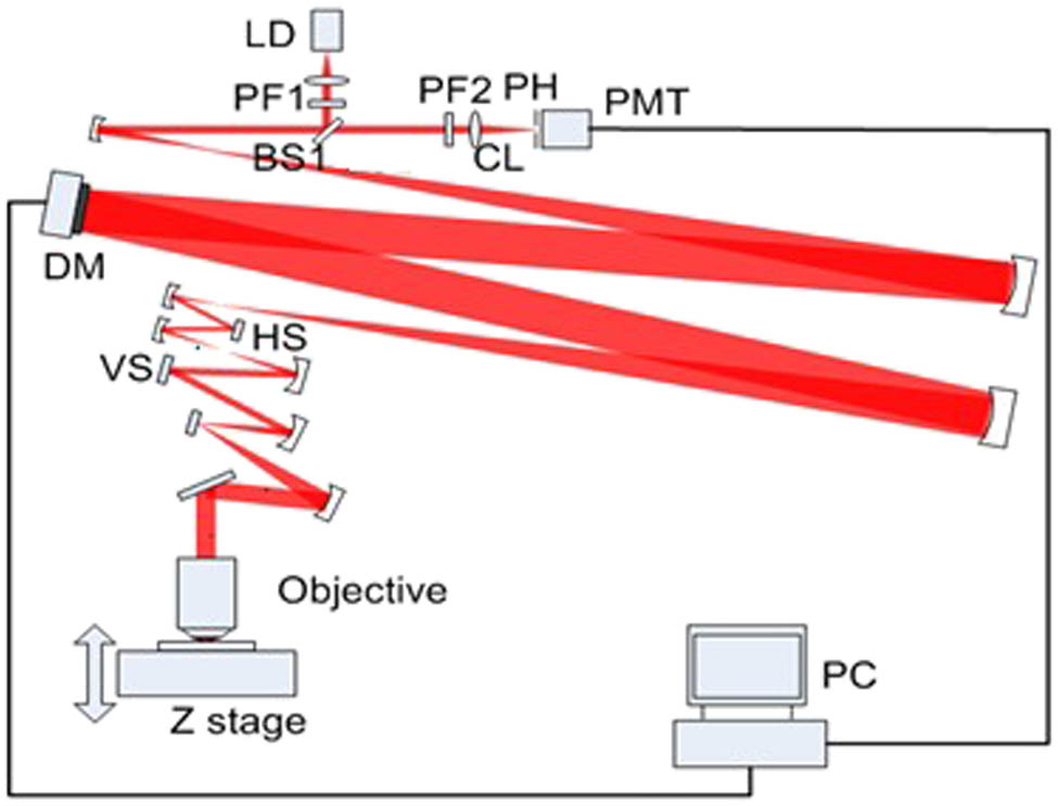
    Fig. 1. Scheme of AO confocal fluorescence microscopy system. BS: beam splitter. PF: bandpass filter. HS: horizontal scanner. VS: vertical scanner. PH: pinhole. CL: collecting lens.
    Fluorescence images without and with AO correction at different depth, and fluorescence signal profiles corresponding to each depth: (A) 80, (B) 100, and (C) 120 μm. The image size is 314*314 microns with a scale bar of 10 μm.
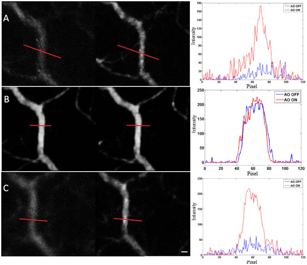
    Fig. 2. Fluorescence images without and with AO correction at different depth, and fluorescence signal profiles corresponding to each depth: (A) 80, (B) 100, and (C) 120 μm. The image size is 314*314 microns with a scale bar of 10 μm.
    Metric value of images during AO correction process in Fig. 2.
    Fig. 3. Metric value of images during AO correction process in Fig. 2.
    Yi He, Zhibin Wang, Yuanyuan Wang, Ling Wei, Xiqi Li, Jinsheng Yang, Guohua Shi, Yudong Zhang. In vivo imaging of mice auricle vessels using adaptive optical confocal fluorescence microscope[J]. Chinese Optics Letters, 2015, 13(11): 111702
    Download Citation